-
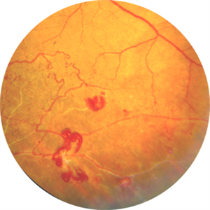
 Cheese Pizza Pie Appearance in CMV Retinitis
Mar 30 2024 by KANWALJEET HARJOT MADAN, M.S. (Ophthalmology); FAICO (Vitreous - Retina)
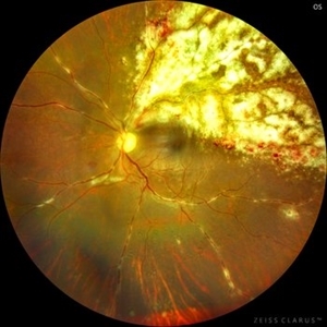
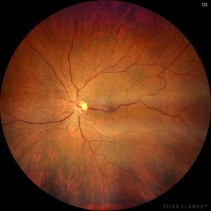
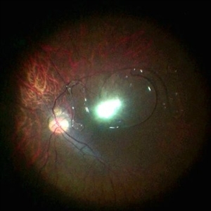
This is Fundus Photograph of left eye of 53 year male depicting an area of Retinal Necrosis with few Retinal Haemorrhages suggestive of CMV Retinitis. Areas of Perivascular Exudation also seen. On investigations, the patient was found to be HIV positive. He was started on Anti Retro Viral treatment after physician opinion.
Photographer: Dr. Kanwaljeet Harjot Madan, Thind Eye Hospital, Jalandhar City (Punjab) INDIA.
Imaging device: Zeiss Fundus Camera
Condition/keywords: AIDS, cytomegalovirus (CMV), retinitis
-
 Combined Central Retinal Vein Occlusion with Branch Retinal Artery Occlusion
Apr 29 2024 by KANWALJEET HARJOT MADAN, M.S. (Ophthalmology); FAICO (Vitreous - Retina)
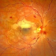
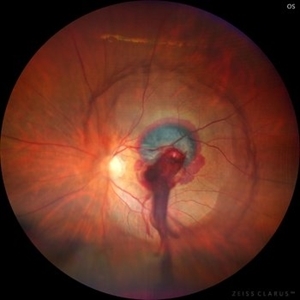
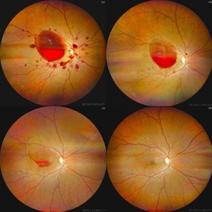
This is fundus photograph of a 46-year male patient who presented with sudden diminution of vision in his right eye (RE) for 3 days. He was hypertensive but non diabetic. On examination, his best corrected vision in RE was 6/12. His left eye (LE) was normal. His fundus examination in RE revealed multiple intra retinal hemorrhages in all quadrants with tortuosity of veins suggestive of central retinal vein occlusion (CRVO) with mild disc edema. An ischemic area was seen superior to fovea suggestive of branch retinal artery occlusion. OCT depicted thickening of inner retinal layers with little evidence of macular edema. Hematological and cardio vascular investigations were done. He had bilateral thickening of intimal and medial walls of carotid arteries. He was under cardiology treatment. His vision improved to 6/6.
Photographer: Dr. Kanwaljeet Harjot Madan, M.S. (Ophthalmologist) Fellow in Vitrous & Retina. Thind Eye Hospital, Jalandhar City. Punjab. India
Condition/keywords: branch retinal artery occlusion (BRAO), central retinal vein occlusion (CRVO)
-
 Combined Central Retinal Vein Occlusion with Branch Retinal Artery Occlusion
Apr 29 2024 by KANWALJEET HARJOT MADAN, M.S. (Ophthalmology); FAICO (Vitreous - Retina)
This is fundus photograph of a 46-year male patient who presented with sudden diminution of vision in his right eye (RE) for 3 days. He was hypertensive but non diabetic. On examination, his best corrected vision in RE was 6/12. His left eye (LE) was normal. His fundus examination in RE revealed multiple intra retinal hemorrhages in all quadrants with tortuosity of veins suggestive of central retinal vein occlusion (CRVO) with mild disc edema. An ischemic area was seen superior to fovea suggestive of branch retinal artery occlusion. OCT depicted thickening of inner retinal layers with little evidence of macular edema. Hematological and cardio vascular investigations were done. He had bilateral thickening of intimal and medial walls of carotid arteries. He was under cardiology treatment. His vision improved to 6/6.
Photographer: Dr. Kanwaljeet Harjot Madan, M.S. (Ophthalmologist) Fellow in Vitrous & Retina. Thind Eye Hospital, Jalandhar City. Punjab. India
Condition/keywords: branch retinal artery occlusion (BRAO), central retinal vein occlusion (CRVO)
-
 Congenital Retinal Macro Vessel
May 15 2024 by KANWALJEET HARJOT MADAN, M.S. (Ophthalmology); FAICO (Vitreous - Retina)
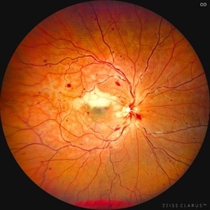
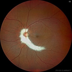
This is the Fundus Picture of a 56 years Female, who had come for Cataract Surgery of her left eye. Her best corrected vision in left eye was 6/12. She was diagnosed to have Congenital Retinal Macro vessel in her left eye on fundus exam. She underwent cataract surgery and vision improved to 6/9. She is kept under regular follow up.
Photographer: Dr. Kanwaljeet Harjot Madan
Condition/keywords: congenital, RETINAL MACROVESSEL
-
 Congenital Retinal Macro Vessel
May 15 2024 by KANWALJEET HARJOT MADAN, M.S. (Ophthalmology); FAICO (Vitreous - Retina)
This is the Fundus Picture of a 56 years Female, who had come for Cataract Surgery of her left eye. Her best corrected vision in left eye was 6/12. She was diagnosed to have Congenital Retinal Macro vessel in her left eye on fundus exam. She underwent cataract surgery and vision improved to 6/9. She is kept under regular follow up.
Photographer: Dr Kanwaljeet Harjot Madan
Condition/keywords: congenital, RETINAL MACROVESSEL
-
 Ruptured Retinal Artery Macroaneurysm
Jun 18 2024 by KANWALJEET HARJOT MADAN, M.S. (Ophthalmology); FAICO (Vitreous - Retina)
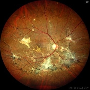
This is a fundus photo depicting ruptured Retinal Artery Macroaneurysm (RAM) in the left eye of a 63 years old female. RAM is an acquired saccular or fusiform dilatation of the retinal arterioles that usually occur within the first three orders of bifurcation. The Superotemporal artery is the most common location. RAM may be asymptomatic or cause a number of complications such as macular edema, serous macular detachment, and hemorrhages.
Photographer: Dr Kanwaljeet Harjot Madan
Condition/keywords: Haemorrhage, macroaneurysm, retinal arteriole
-
 Myelinated Nerve Fibres
Jul 15 2024 by KANWALJEET HARJOT MADAN, M.S. (Ophthalmology); FAICO (Vitreous - Retina)
This is Fundus Photograph of a Female Patient aged 53 years. She had Myelinated Nerve Fibres with Lamellar Macular Hole in left eye. She underwent surgery for Lamellar Macular Hole and her vision improved from 20/70 to 20/30.
Photographer: Dr. Kanwaljeet Harjot Madan, Thind Eye Hospital, Jalandhar City. Punjab. India.
Imaging device: Ziess Clarus
Condition/keywords: fundus photograph, lamellar macular hole, Myelinated Nerve Fibres
-
 Pseudoxanthoma Elasticum Associated Angioid Streaks
Aug 18 2024 by KANWALJEET HARJOT MADAN, M.S. (Ophthalmology); FAICO (Vitreous - Retina)
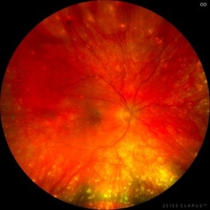
This is fundus photograph of a young 31 years male patient depicting Angioid streaks emanating from optic nerve towards the periphery and subretinal fibrosis. There is peau de orange appearance temporal to fovea with Salmon Spots in periphery. He was diagnosed to have Pseudoxanthoma Elasticum.
Photographer: Dr. Kanwaljeet Harjot Madan, M.S. (Ophthalmologist) Fellow in Vitrous & Retina. Thind Eye Hospital, Jalandhar City. Punjab. India
Imaging device: Zeiss Clarus
Condition/keywords: Angioid Streaks, fundus photograph, pseudoxanthoma elasticum (PXE)
-
 Valsalva Retinopathy
Sep 10 2024 by KANWALJEET HARJOT MADAN, M.S. (Ophthalmology); FAICO (Vitreous - Retina)
These are fundus pictures of a young 32 years male who presented with sudden decrease in vision in right eye after lifting heavy weights. His vision in right eye was hand movements close to face. He had no systemic history or any history of trauma. He was diagnosed to have Valsalva Retinopathy. His hematological investigations were normal. He refused any active intervention. He was kept under observation and his vision improved to 20/20 within 4 weeks.
Photographer: Dr. Kanwaljeet Harjot Madan, M.S. (Ophthalmologist) Fellow in Vitrous & Retina. Thind Eye Hospital, Jalandhar City. Punjab. India
Imaging device: Zeiss Clarus
Condition/keywords: valsalva retinopathy
-
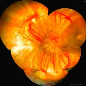
 Sunset Glow Fundus
Nov 11 2024 by KANWALJEET HARJOT MADAN, M.S. (Ophthalmology); FAICO (Vitreous - Retina)
This is fundus picture of right eye of a 15 year-old female patient depicting convalescent phase of Vogt Koyanagi Harada Syndrome. This phase occurs few weeks to few months after the acute stage of the disease. Fundus has a typical bright orange appearance, also known as SUNSET GLOW, along with appearance of DALEN FUCHS nodules.
Photographer: Dr. Kanwaljeet Harjot Madan, M.S. (Ophthalmologist) Fellow in Vitrous & Retina. Thind Eye Hospital, Jalandhar City. Punjab. India
Imaging device: Zeiss Clarus
Condition/keywords: Dalen-Fuchs nodules, Sunset Glow Fundus, VOGT KOYANAGI HARADA
-
 Benign Familial Fleck Retina
Dec 2 2024 by KANWALJEET HARJOT MADAN, M.S. (Ophthalmology); FAICO (Vitreous - Retina)
This is fundus picture of a 21 year old female patient who had come for refractive surgery consultation. Her best corrected vision in both eyes was 20/20. She had myopic astigmatism in both eyes. Fundus exam revealed presence of multiple yellowish white flecks spread throughout retina sparing macular area in both eyes. Her color vision was normal. Electroretinogram and electrooculogram were normal. She gave no history of night blindness. A diagnosis of Benign Familial Fleck Retina was made. She was also advised ocular exam of her parents and elder brother which was normal.
Photographer: Dr. Kanwaljeet Harjot Madan, M.S. (Ophthalmologist) Fellow in Vitrous & Retina. Thind Eye Hospital, Jalandhar City. Punjab. India
Imaging device: Zeiss Clarus
Condition/keywords: Benign familial fleck retina, Night Blindness
-
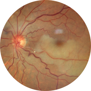
 Comets in the Eye (Retinopathy of Prematurity)
Apr 8 2025 by KANWALJEET HARJOT MADAN, M.S. (Ophthalmology); FAICO (Vitreous - Retina)
This is the fundus picture of right eye (RE) of a 4 years female child presented with outward deviation of right eye. Her parents also complained of diminution of vision in both eyes. On examination, her best corrected vision in RE was hand movements close to face and was 20/80 in LE. Posterior segment exam revealed presence of macular scar in RE and presence of dry retinal fold with dragging of retinal vessels. LE fundus revealed presence of nasal drag of optic disc. Parents gave history of untreated ROP as an infant. Retinopathy of Prematurity (ROP) is a Vaso proliferative disorder of Retina occurring in premature infants. Advances in neonatal care and ROP treatment has led these babies to live longer with this disease.
Photographer: Dr. Kanwaljeet Harjot Madan, Thind Eye Hospital, Jalandhar City (Punjab) INDIA.
Imaging device: Zeiss Fundus Camera
Condition/keywords: Retinopathy of Prematurity
-
 Comets in the Eye (Retinopathy of Prematurity)
Apr 8 2025 by KANWALJEET HARJOT MADAN, M.S. (Ophthalmology); FAICO (Vitreous - Retina)
This is the fundus picture of right eye (RE) of a 4 years female child presented with outward deviation of right eye. Her parents also complained of diminution of vision in both eyes. On examination, her best corrected vision in RE was hand movements close to face and was 20/80 in LE. Posterior segment exam revealed presence of macular scar in RE and presence of dry retinal fold with dragging of retinal vessels. LE fundus revealed presence of nasal drag of optic disc. Parents gave history of untreated ROP as an infant. Retinopathy of Prematurity (ROP) is a Vaso proliferative disorder of Retina occurring in premature infants. Advances in neonatal care and ROP treatment has led these babies to live longer with this disease.
Photographer: Dr. Kanwaljeet Harjot Madan, Thind Eye Hospital, Jalandhar City (Punjab) INDIA.
Imaging device: Zeiss Fundus Camera
Condition/keywords: Retinopathy of Prematurity, Vaso proliferative disorder
-
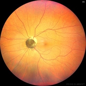 Optic Nerve Melanocytoma
May 4 2025 by KANWALJEET HARJOT MADAN, M.S. (Ophthalmology); FAICO (Vitreous - Retina)
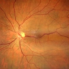
This is a fundus picture of a young 42-year male who visited for a routine eye exam. His BCVA was 20/20 in both eyes. Anterior segment examination was normal. His left eye showed grey-black pigmentation at the infero-nasal margin of the optic disc. Fundus of the right eye was normal. The patient was diagnosed to have optic disc melanocytoma on multimodal imaging and was advised regular follow-up. Optic nerve melanocytoma is typically a benign tumor made up of melanocytes and melanin. It can grow, but rarely transforms into a malignancy. Patients with Optic Nerve Melanocytoma should be periodically examined for evidence of growth, loss of visual field and optic nerve compression.
Photographer: Dr. Kanwaljeet Harjot Madan, Thind Eye Hospital, Jalandhar City (Punjab) INDIA.
Imaging device: Zeiss Fundus Camera
Condition/keywords: melanocytoma, melanoma, optic nerve
-
 Aggressive Posterior Retinopathy of Prematurity (APROP)
May 16 2025 by KANWALJEET HARJOT MADAN, M.S. (Ophthalmology); FAICO (Vitreous - Retina)
This is the fundus picture of right eye of a premature neonate depicting Aggressive Posterior Retinopathy of Prematurity (APROP). It is a severe rapidly progressing form of retinopathy that can lead to vision loss and blindness. It requires prompt diagnosis and treatment in the form of anti-VEGF agents and laser photocoagulation.
Photographer: Dr. Kanwaljeet Harjot Madan, Thind Eye Hospital, Jalandhar City (Punjab) INDIA.
Imaging device: Zeiss Clarus
Condition/keywords: Oxygen Exposure, retinopathy of prematurity (ROP)
-
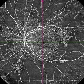 Proliferative Diabetic Retinopathy
May 29 2025 by KANWALJEET HARJOT MADAN, M.S. (Ophthalmology); FAICO (Vitreous - Retina)
This is widefield optic coherence tomography angiography (WF-OCTA) picture of LE of a diabetic patient. This patient had Proliferative Diabetic Retinopathy and depicts large areas of capillary non perfusion with neovascularization elsewhere.
Photographer: Dr. Kanwaljeet Harjot Madan, Thind Eye Hospital, Jalandhar City (Punjab) INDIA.
Imaging device: Widefield Optic Coherence Tomography Angiography (WF-OCTA).
Condition/keywords: OCTA, proliferative diabetic retinopathy (PDR), ultra-wide field imaging
-
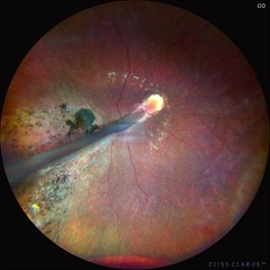
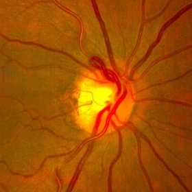 Prepapillary Vascular Loop
Jul 4 2025 by KANWALJEET HARJOT MADAN, M.S. (Ophthalmology); FAICO (Vitreous - Retina)
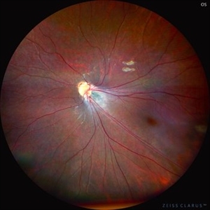
This is the fundus picture of right eye of a young 32 years female depicting pre papillary vascular loop. A prepapillary vascular loop is a congenital anomaly of the optic disc that presents as an elevated and twisted bundle of vessels projecting into the vitreous cavity. It is a benign condition, usually unilateral but can be bilateral. It is asymptomatic and discovered during routine eye examination. This anomaly can sometimes cause complications like branch retinal artery occlusion, vitreous hemorrhage, or sub retinal hemorrhage.
Photographer: Dr. Kanwaljeet Harjot Madan, Thind Eye Hospital, Jalandhar City (Punjab) INDIA.
Imaging device: Zeiss Fundus Camera
Condition/keywords: branch retinal artery occlusion (BRAO), optic disc, Prepapillary Vascular Loop, SUB RETINAL HEMORRHAGE, Vitreous hemorrhage
-
 Desert Rose
Aug 4 2025 by KANWALJEET HARJOT MADAN, M.S. (Ophthalmology); FAICO (Vitreous - Retina)
This is fundus picture of left eye of a 54 years male depicting Supero Temporal Branch Retinal Vein Occlusion with collaterals and neo vascularization elsewhere.
Photographer: Dr. Kanwaljeet Harjot Madan, Thind Eye Hospital, Jalandhar City (Punjab). INDIA.
Imaging device: Zeiss Fundus Camera
Condition/keywords: Neo Vascularization, Supero Temporal Branch Retinal Vein Occlusion
-
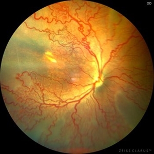
 Albinotic Fundus
Aug 18 2025 by KANWALJEET HARJOT MADAN, M.S. (Ophthalmology); FAICO (Vitreous - Retina)
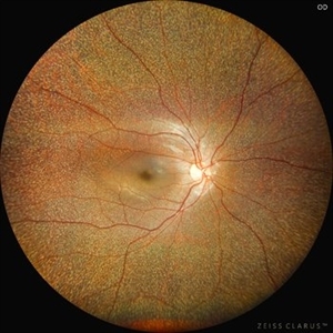
This is ultrawide field montage image of a 40 year-old male depicting Hypopigmented Albinotic Fundus. Arborizing network of choroidal vasculature is prominent. Multiple vortex vein ampullae near equator and posterior pole are clearly visible.
Photographer: Dr. Kanwaljeet Harjot Madan, Thind Eye Hospital, Jalandhar City (Punjab). INDIA.
Imaging device: Zeiss Fundus Camera
Condition/keywords: Albinotic Fundus, choroidal vasculature, vortex vein
-
 Hollenhorst Plaque
Sep 2 2025 by KANWALJEET HARJOT MADAN, M.S. (Ophthalmology); FAICO (Vitreous - Retina)
A 64 year-old male presented with sudden decrease in vision in LE for 1 week. His BCVA in LE was 20/200. Fundus exam revealed presence of whitish ischemic area in macula superior to fovea suggestive of branch retinal artery occlusion. A bright tiny refractile cholesterol embolus (Hollenhorst plaque) was visible in retinal artery. The patient was advised cardiology consultation.
Photographer: Dr. Kanwaljeet Harjot Madan, Thind Eye Hospital, Jalandhar City (Punjab). INDIA.
Imaging device: Zeiss Fundus Camera
Condition/keywords: branch retinal artery occlusion (BRAO), hollenhorst plaque
-
 Dislocated ACIOL
Oct 23 2025 by KANWALJEET HARJOT MADAN, M.S. (Ophthalmology); FAICO (Vitreous - Retina)
This is intraoperative image of a young male who presented with sudden diminution of vision in RE after Trauma. Fundus exam revealed presence of dislocated anterior chamber IOL in Vitreous Cavity.
Photographer: Dr. Kanwaljeet Harjot Madan, Thind Eye Hospital, Jalandhar City (Punjab). INDIA.
Imaging device: Zeiss Fundus Camera
Condition/keywords: dislocated anterior chamber intraocular lens (ACIOL)
-
 Leber’s Miliary Aneurysm
Dec 12 2025 by KANWALJEET HARJOT MADAN, M.S. (Ophthalmology); FAICO (Vitreous - Retina)
A 34 year-old male presented with decrease vision in right eye for 3 months. Anterior segment exam was normal. Fundus exam in RE revealed presence of macular edema which was evident on OCT. Multiple retinal vascular aneurysmal dilatations with telangiectasia of the retina blood vessels noted superiorly which was evident on FFA. These aneurysms were multiple, tiny and leaky on FFA. He was diagnosed to have Leber’s miliary aneurysms. It is a rare, typically unilateral eye condition, often seen in young males, characterized by multiple tiny, leaky aneurysms in the retinal blood vessels, leading to deposits of hard exudates and potential vision loss, especially if it affects the macula. It is considered a milder form of Coats' disease.
Photographer: Dr. Kanwaljeet Harjot Madan, Thind Eye Hospital, Jalandhar City (Punjab) INDIA.
Imaging device: Zeiss Fundus Camera
Condition/keywords: FFA, Leber's miliary aneurysm
-
 Best Vitelliform Macular Dystrophy
Dec 24 2025 by KANWALJEET HARJOT MADAN, M.S. (Ophthalmology); FAICO (Vitreous - Retina)
This is Fundus Autofluorescence (FAF) image of LE of a 7 year-old boy. He was diagnosed to have retinal lesions in BE elsewhere. Fundus exam revealed presence of egg yolk macular lesions in BE which depicted intense hyper autofluorescence. He was diagnosed to have Best Vitelliform Macular Dystrophy. It is an autosomal dominant condition caused by mutations in BEST 1 Gene and leads to accumulation of lipofuscin in subretinal space.
Photographer: Dr. Kanwaljeet Harjot Madan, Thind Eye Hospital, Jalandhar City (Punjab) INDIA.
Imaging device: Zeiss Fundus Camera
Condition/keywords: Best vitelliform macular dystrophy (BVMD)
-
 Retinal Astrocytoma
Jan 28 2026 by KANWALJEET HARJOT MADAN, M.S. (Ophthalmology); FAICO (Vitreous - Retina)
This is the fundus image of LE of a 16 year-old male depicting presence of Retinal Astrocytic Hamartoma in peripapillary region. Few depigmented areas of retinal pigment epithelium can be seen infero-temporally. He had associated Tuberous Sclerosis. Retinal Astrocytoma is a benign glial cell tumor, often asymptomatic tumor affecting 40-50% of patients with Tuberous Sclerosis Complex (TSC).
Photographer: Dr. Kanwaljeet Harjot Madan, Thind Eye Hospital, Jalandhar City (Punjab) INDIA.
Imaging device: Zeiss Fundus Camera
Condition/keywords: astrocytoma, tuberous sclerosis
A project from the American Society of Retina Specialists