 Initializing download.
-
 By Anand Temkar
Co-author(s): Dr.Manish Nagpal - Retina Foundation, Ahmedabad - Uploaded on Nov 29, 2024.
- Last modified by Anand Temkar on Dec 2, 2024.
- Rating
- Appears in
- Choroidal Hemangioma
- Condition/keywords
- FLUORESCEIN ANGIOGRAPHY, indocyanine green (ICG) angiography, Choroidal Hemangioma
- Photographer
- Dr.Anand Temkar- Retina Foundation, Ahmedabad
- Imaging device
-
Scanning laser ophthalmoscope
Mirante - Description
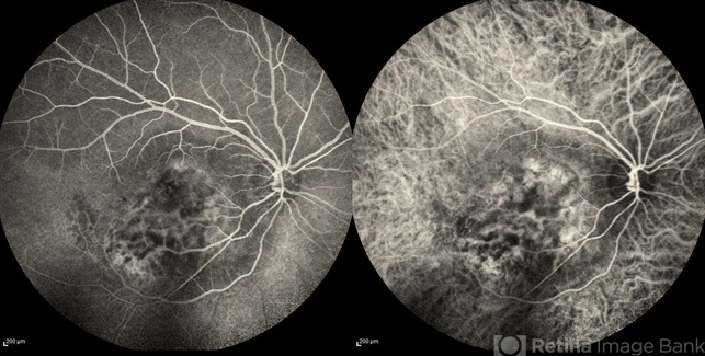
- Right eye Fluorescein and Indocyanine green angiography of a 42 year old male in case of Choroidal hemangioma. Choroidal hemangioma have a unique pattern of circulation where the large blood vessels produce a “COARSE VASCULAR PATTERN.” Fluorescein angiography of circumscribed choroidal hemangiomas typically reveals very early hyperfluorescence of larger-caliber choroidal blood vessels either before or simultaneously with the initial filling of the retinal arterioles. Indocyanine green angiography typically shows filling of the intralesional vascular channels, intense hypercyanescence of the lesion by the intermediate frames (peaks around 3-4 minutes) and late washout of the central portion of the lesion.
")


")
")
")
")

")