-
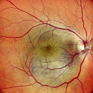 Right Eye Color Photo in Case of Choroidal Hemangioma
Nov 29 2024 by Anand Temkar
Right eye color photo of a 42 year old male in case of choroidal hemangioma. We can see the reddish-orange, round to oval choroidal tumor located completely in the posterior half of the fundus.
Photographer: Dr.Anand Temkar- Retina Foundation, Ahmedabad
Imaging device: Mirante
Condition/keywords: Choroidal Hemangioma
-
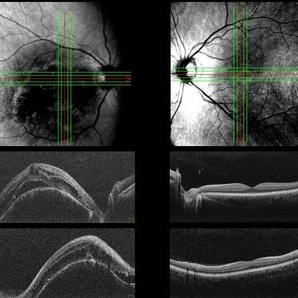 Both Eyes OCT in Case of Right Eye Choroidal Hemangioma
Nov 29 2024 by Anand Temkar
BE OCT of a 42 year old male, showing the elevation of the right eye retina along with the cystic spaces and subretinal fluid.
Photographer: Dr.Anand Temkar- Retina Foundation, Ahmedabad
Imaging device: Mirante
Condition/keywords: OCT
-
 Fluorescein and Indocyanine Green Angiography in Right Eye in Case of Choroidal Hemangioma
Nov 29 2024 by Anand Temkar
Right eye Fluorescein and Indocyanine green angiography of a 42 year old male in case of Choroidal hemangioma. Choroidal hemangioma have a unique pattern of circulation where the large blood vessels produce a “COARSE VASCULAR PATTERN.” Fluorescein angiography of circumscribed choroidal hemangiomas typically reveals very early hyperfluorescence of larger-caliber choroidal blood vessels either before or simultaneously with the initial filling of the retinal arterioles. Indocyanine green angiography typically shows filling of the intralesional vascular channels, intense hypercyanescence of the lesion by the intermediate frames (peaks around 3-4 minutes) and late washout of the central portion of the lesion.
Photographer: Dr.Anand Temkar- Retina Foundation, Ahmedabad
Imaging device: Mirante
Condition/keywords: Choroidal Hemangioma, FLUORESCEIN ANGIOGRAPHY, indocyanine green (ICG) angiography
-
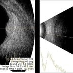 USG in Case of Choroidal Hemangioma With Associated Exudative RD
Nov 29 2024 by Anand Temkar
USG of right eye of a 42 year old male, showing us the BRIGHT choroidal hemangioma. This is because they are a mass of relatively large and well-formed blood vessels. Each blood vessel reflects sound waves producing characteristically intense reflections or moderately “MODERATELY HIGH INTERNAL REFLECTIVITY” from within the hemangioma tumor. Moderately high internal reflectivity is an important diagnostic characteristic.
Photographer: Dr.Anand Temkar- Retina Foundation, Ahmedabad
Imaging device: Mirante
Condition/keywords: B scan ultrasound, Choroidal Hemangioma
A project from the American Society of Retina Specialists