 Initializing download.
-
 By KRISHNENDU NANDI, MS
Netralayam Superspeciality Eye Care Centre, Kolkata, India - Uploaded on Feb 6, 2024.
- Last modified by Joshua Friedman on Feb 6, 2024.
- Rating
- Appears in
- Miscellaneous
- Condition/keywords
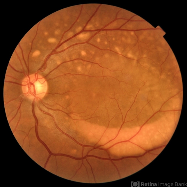
- Best vitelliform macular dystrophy (BVMD), Multifocal
- Photographer
- Dr. Krishnendu Nandi
- Imaging device
- Fundus camera
- Description
- A 38-year-old male presented with gradual dimness of vision in both eyes for last 3 months. Best corrected visual acuity was 6/24, N8 in both eyes. Colour fundus photograph showed multiple orangish yellow sub retinal lesions on the posterior pole extending beyond arcades. Macular thickening also noted. OCT line scan through the fovea showed thickened ellipsoid zone and it was separated from the RPE by optically clear space.


