-
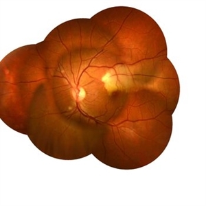
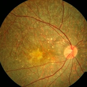
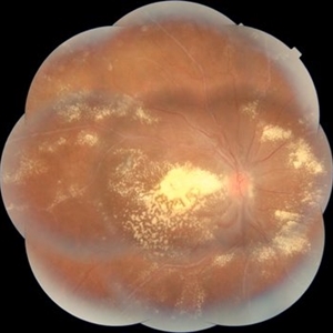
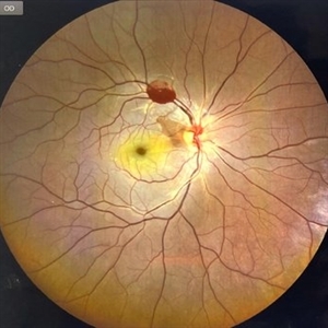
 RAM With Garland of Hard Exudate
Mar 3 2020 by KRISHNENDU NANDI, MS
Fundus Photo of left eye of 75-year-old female with retinal artery macroaneurysm at superior quadrant with garland like hard exudates.
Photographer: KRISHNENDU NANDI
Imaging device: Topcon
Condition/keywords: hard exudates, retinal arterial macroaneurysm
-
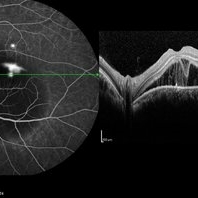 Fibrin Dipping Sign in OCT in Smokestack CSCR Leak
Aug 27 2020 by KRISHNENDU NANDI, MS
Image of a 42-year-old male showing smokestack CSCR leak in DFA with subretinal fibrin generates a dipping morphological pattern on OCT.
Photographer: Dr. Krishnendu Nandi
Condition/keywords: central serous chorioretinopathy (CSCR), fibrin
-
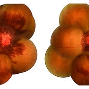
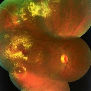 Coats' Disease
Oct 31 2020 by KRISHNENDU NANDI, MS
Fundus image of Right eye of a 12-year-old boy presented with dimness of vision for 6 months. Image showed lot exudation along the telengiectatic vessels at periphery. Cystoid macular edema also noted.
Photographer: Krishnendu Nandi, Netralayam Eye Hospital, Kolkata, India
Condition/keywords: Coats' disease, retinal telangiectasia
-
 ST BRAO
Nov 5 2020 by KRISHNENDU NANDI, MS
A 26-year-old male with typhoid fever presented with ST BRAO in left eye.
Photographer: Krishnendu Nandi
Condition/keywords: branch retinal artery occlusion (BRAO), typhoid fever
-
 Bietti's Crystalline Dystrophy with CNVM
Dec 14 2020 by KRISHNENDU NANDI, MS
Fundus picture of right eye of 29-year-old female with Bietti's crystalline dystrophy with CNVM.
Photographer: Krishnendu Nandi, B B Eye Foundation VIP, Kolkata, India
Condition/keywords: Bietti's crystalline dystrophy, choroidal neovascular membrane (CNVM)
-
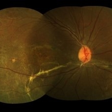 Spontaneously Attached Retina
Jan 15 2021 by KRISHNENDU NANDI, MS
Fundus photograph of 26-year-old man with BCVA 6/60, N24, showed spontaneous retinal reattachment with multiple retinal cyst at temporal quadrant. Subretinal gliosis resemble a mustache.
Photographer: Krishnendu Nandi, Netralayam Eye Care Centre
Condition/keywords: spontaneous retinal reattachment
-
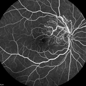 Pre-Papillary Vascular Loop
Jul 8 2021 by KRISHNENDU NANDI, MS
DFA picture of 42-year-old male presented with dimness of vision in left eye for 3 months. DFA showed pre papillary vascular loop.
Photographer: Krishnendu Nandi, Netralayam Eye Care Centre, Kolkata, India
Condition/keywords: congenital prepapillary vascular loop
-
 Anemic Retinopathy in a Young Female
Jan 15 2022 by KRISHNENDU NANDI, MS
Fundus photograph of a 29-year-old female presented with retinal hemorrhages in both eyes with decrease in vision for 1 month. Hemoglobin level was 5.9gm/dl, suggestive of anemic retinopathy in both eyes.
Photographer: Krishnendu Nandi, Netralayam Eye Care Centre, Kolkata, India
Imaging device: Topcon
Condition/keywords: anaemic retinopathy, anemic retinopathy, retinal hemorrhage
-
 IRVAN
IRVAN
Jan 20 2022 by KRISHNENDU NANDI, MS
A 29-year-old female presented with decreased vision in both eyes. OD: CF 4 meters, OS: 20/60. Fundus Photo of right eye showed hard exudates at macula scattered throughout posterior pole. Optic Disc is mildly swollen in both eyes. Multiple saccular aneurysmal dilatation was noted along the retinal tree in both eyes. Features of Retinal vasculitis also noted in both eyes; suggestive of IRVAN syndrome.
Photographer: Krishnendu Nandi, Netralayam Eye Care Centre, Kolkata, India
Condition/keywords: IRVAN Syndrome, retinal vasculitis
-
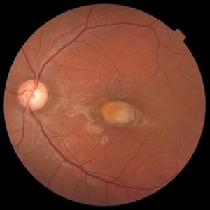 Torpedo Maculopathy
Mar 8 2022 by KRISHNENDU NANDI, MS
Fundus Photograph of a 25 year young male came with complain of distortion of vision in left eye for last few months. BCVA was 6/9, N6 in left eye. Retinal examination revealed torpedo maculopathy in left eye.
Photographer: Krishnendu Nandi, Netralayam Eye Care Centre, Kolkata, India
Condition/keywords: torpedo maculopathy, Young Male
-
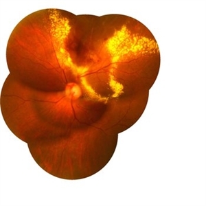
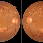 Multifocal Best Disease
Aug 3 2023 by KRISHNENDU NANDI, MS
A 38-year-old male presented with gradual dimness of vision in both eyes for last 3 months. Best corrected visual acuity was 6/24, N8 in both eyes. Colour fundus photograph showed multiple orangish yellow sub retinal lesions on the posterior pole extending beyond arcades. Macular thickening also noted. Fundus auto-fluorescence showed multiple white hyper auto-fluorescence suggestive of RPE dysfunction. OCT line scan through the fovea showed thickened ellipsoid zone and it was separated from the RPE by optically clear space
Photographer: Krishnendu Nandi
Condition/keywords: Best Disease, Multifocal, Young Male
-
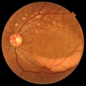 Multifocal Best Disease
Feb 6 2024 by KRISHNENDU NANDI, MS
A 38-year-old male presented with gradual dimness of vision in both eyes for last 3 months. Best corrected visual acuity was 6/24, N8 in both eyes. Colour fundus photograph showed multiple orangish yellow sub retinal lesions on the posterior pole extending beyond arcades. Macular thickening also noted. OCT line scan through the fovea showed thickened ellipsoid zone and it was separated from the RPE by optically clear space.
Photographer: Dr. Krishnendu Nandi
Condition/keywords: Best vitelliform macular dystrophy (BVMD), Multifocal
-
 Traumatic CRAO with Cilioretinal Artery Sparing
Sep 10 2024 by KRISHNENDU NANDI, MS
A 25 year-old male presented with dimness of vision in the right eye for the last 3 days following blunt trauma. The BCVA of the right eye was CF close to the face and left eye was 6/6, N6 in Snellen’s chart. On examination the retina showed CRAO with cherry red spot and sparing of cilioretinal artery circulation. Traumatic subhyaloid haemorrhage also noted at supero-temporal arcade.
Photographer: Dr Krishnendu Nandi
Condition/keywords: CRAO, subhyaloid hemorrhage, Trauma
A project from the American Society of Retina Specialists