 Initializing download.
-
 By McGill University Health Centre
The MUHC-McGill University
Co-author(s): Miguel N. Burnier, Paulina García de Alba Graue, McGill University Health Center-McGill University Ocular Pathology & Translational Research Laboratory - Uploaded on Dec 10, 2020.
- Last modified by Caroline Bozell on Dec 11, 2020.
- Rating
- Appears in
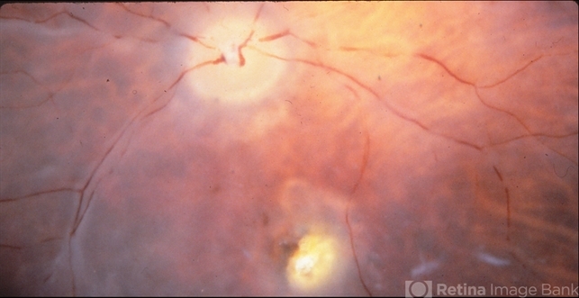
- Best Vitelliform Macular Dystrophy
- Condition/keywords
- Best vitelliform macular dystrophy (BVMD), fundus photograph
- Imaging device
- Fundus camera
- Description
- Postmortem eyes from 101-year-old female. Past clinical history includes a poor vision for many years due to macular degeneration. The last visual acuity test recorded 6/15 OD and 6/6 OS. IOP 14 and 18 torr OS. Histopathology: Disclosed and yellow 2x2mm macular lesion. Microscopic examination: elevated placoid macular lesion with overlying pigment granules. Electron microscopy examination: pigment granules with abundant lipofuscin and melanolysosomes, photoreceptor cells markedly attenuated (less degenerated at the periphery) Numerous calcified drusen throughout the retina particularly in the posterior pole. RPE lipofuscin content is elevated in Best’s dystrophy. The extractability of the PRE lipofuscin fluorophores is reduced (it is normal during senescence). The defect in Best’s dystrophy accelerates this age related change in lipofuscin.
")
")
")


---thumb.JPG/image-square;max$79,0.ImageHandler "Welding arc maculopathy")


---thumb.jpg/image-square;max$79,0.ImageHandler "Normal Fundus Photo")