Search results (14 results)
-
 From Ora to Ora
Aug 26 2024 by Nassim Alejandro Abreu Arbaje, MD
Ultra-wide field OCT angiography of a 39 year-old healthy male. The photo attempts to explore retinal vasculature up to the ora serrata.
Photographer: Johel Arrieta, TowardPi
Imaging device: TowardPi BMizar 400khz
Condition/keywords: OCT Angiography, OCTA, ultra-wide field imaging
-
 Kissing Serous Choroidal Detachment
Mar 8 2023 by Annaka Gooding
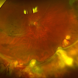
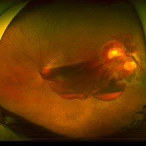
Ultra-widefield fundus photograph of a 73 year old male with a Kissing serous choroidal detachment affecting his right eye. Patient presented at the office following a XEN implant and his vision was sc20/100 PH20/50+1. The physician recommended to start Prednisone treatment.
Photographer: Annaka Gooding
Imaging device: Optos California
Condition/keywords: fundus photography, Kissing Serous Choroidal Detachment, Optos, Right Eye, ultra-wide field imaging
-
 Methotrexate Bubble following Intravitreal Injection for PVR
Sep 21 2022 by Zach Seim
Ultra-widefield fundus photograph of an 81 year old female with a Methotrexate bubble following an Intravitreal Injection for Proliferative Vitreoretinopathy. Patient has been presenting to the office for two week interval Methotrexate injections in her left eye. The image was taken prior to her eighth injection which revealed a residual Methotrexate bubble in her inferior retinal image. This patient was seeing "lots" of floaters, as well as having visual acuity of cc20/400 cc20/200 PH.
Photographer: Zach Seim
Imaging device: OPTOS California
Condition/keywords: bubble, fundus photograph, fundus photography, intravitreal injection, left eye, methotrexate, nasal retina, Optos, proliferative vitreoretinopathy (PVR), pseudocolor, ultra-wide field imaging
-
 Ocular Albinism
Dec 11 2021 by Luis Daniel Gutierrez, MD
Optos images of 6-year-old female patient with type 1 oculocutaneous albinism.
Photographer: Luis Daniel Gutierrez García, Hospital Fundación Nuestra Señora de la Luz, Ciudad de México.
Imaging device: Optos
Condition/keywords: Albinism, ocular albinism, Optos, ultra-wide field imaging
-
 Choroidal Detachment
Jan 17 2022 by Logan ryzenga
Left ultra-wide field photograph of an 81-year old female with a choroidal detachment affecting her left eye. Patient had a stent placed November, 2021 and following the procedure she complains of variable blurred vision and severe constricted visual fields. She presented at our office with flashes a month prior but without pain or floaters.
Photographer: Logan Ryzenga
Imaging device: Optos California
Condition/keywords: choroidal detachment, fundus photograph, left eye, Optos, pseudocolor, superior retina, ultra-wide field imaging
-
 Ultra-Widefield Fundus Image of Coats' Disease with Exudative Retinal Detachment
Dec 22 2020 by Kushal S Delhiwala, MBBS, MS, FMRF,FICO, FAICO
Ultra-widefield fundus image of right eye showing Coats' disease with exudative retinal detachment (stage 3) in a 4 year old male complaining of squinting right eye. Telangiectatic vessels prominent superotemporal periphery.
Photographer: Kushal Delhiwala, Netralaya superspeciality eye hospital, Ahmedabad, Gujarat,India
Imaging device: Optos Daytona
Condition/keywords: bullous retinal detachment, Coats' disease, exudative detachment, ultra-wide field imaging
-
 Hydrogel Implant Intrusion
May 5 2020 by Geovanni Jassiel Rios, MD
Ultra-wide field fundus photograph of the right eye with reattached retina. We can observe retinal hydrogel implant intrusion at the inferior retina
Photographer: Ericka , Hospital de la Luz
Condition/keywords: hydrogel implant intrusion, ultra-wide field imaging
-
 Morning Glory Syndrome
Jan 6 2020 by Olivia Rainey
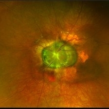
Ultra-wide field pseudocolor image of a 23-month-old male with morning glory syndrome affecting his left eye. Patient presented with esotropia affecting his left eye and strabismic amblyopia affecting both eyes. He could fix and follow on exam and his medical history was unremarkable.
Photographer: Olivia Rainey
Imaging device: Optos California
Condition/keywords: esotropia, left eye, macular, Morning Glory Syndrome, Optos, strabismic amblyopia, ultra-wide field imaging
-
 Coats' Disease
Jul 16 2019 by Kim Barrett
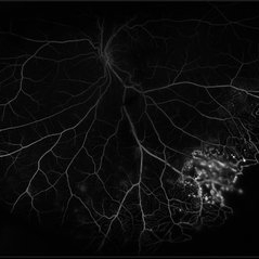
Ultra-wide field fluorescein angiogram of a 23-year-old male with Coats' disease, presented with distorted vision affecting his left eye. He reported seeing flashes and floaters since January of 2019, but the flashes had resolved. He was treated with Intravitreal Preservative Free Triamcinolone in the office and scheduled for PRP laser in the near future.
Photographer: Kim Barrett
Imaging device: Optos
Condition/keywords: Coats' disease, fluorescein angiogram (FA), fluorescein leakage, inferior retina, ischemia, left eye, Optos, ultra-wide field imaging
-
 Morning Glory Syndrome
Jun 19 2019 by Olivia Rainey
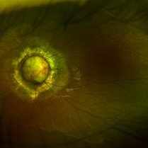
Ultra-wide field pseudocolor image of an 10-year-old girl with Morning Glory Syndrome affecting her left eye. Patient is able to count fingers at 4 feet.
Photographer: Olivia Rainey
Imaging device: Optos
Condition/keywords: left eye, Morning Glory Syndrome, Optos, pseudocolor, ultra-wide field imaging
-
 Penetrating Trauma with Retinal Detachment
Apr 30 2019 by Olivia Rainey
Ultra-wide field pseudocolor image of a 39-year-old female with penetrating trauma resulting in a retinal detachment with an intraretinal hemorrhage affecting the left eye. Patient was struck with a champagne glass in October of 2018, which lacerated the eyelid and globe. Patient was "seeing red" when she first came to the office and after multiple surgeries she was seeing 20/20 at her last check in April 2019.
Photographer: Olivia Rainey
Imaging device: Optos
Condition/keywords: hemorrhage, left eye, Optos, penetrating trauma, ruptured globe, ultra-wide field imaging
-
 Choroidal Melanoma
Jan 30 2019 by Karen Panzegrau
Ultra-wide field optos image of a 27-year-old male patient who presented with loss of vision for about 6-8 weeks. Previous choroidal nevus seen. Recommended annual monitoring. No exam for since 10/2014. Brachytherapy vs enucleation was discussed. Brachytherapy was decided as treatment. Full metastatic work up is being performed.
Photographer: Karen Panzegrau
Imaging device: Optos
Condition/keywords: choroidal nevus, exudative retinal detachment, malignant neoplasm of eye, Optos, ultra-wide field imaging
-
 Multiple Myeloma with Cytomegalovirus Retinitis
Apr 5 2018 by Kim Barrett
Ultra-wide field fluorescein angiogram of a 77-year-old male with multiple myeloma. Patient's angiogram presented significant peripheral retinal ischemia and cystoid macular edema. Patient tested positive for polymerase chain reaction, confirming cytomegalovirus retinitis. Patient is being treated with intravitreal ganciclovir and his current vision is 20/200.
Photographer: Kim Barrett, COA
Imaging device: Optos
Condition/keywords: cystoid macular edema (CME), fluorescein angiogram (FA), fluorescein leakage, intravitreal ganciclovir, myeloma, peripheral ischemia, positive polymerase chain reaction (PCR), ultra-wide field imaging
-
 UWF of Retinal Detachment Corrected with Scleral Buckle
Aug 29 2017 by Carolyn Daley
An ultra wide field fundus photograph of a 57-year-old male who has a past history of retinal detachment corrected with scleral buckle and three treated retinal tears.
Photographer: Carolyn Daley
Imaging device: Optos Imaging
Condition/keywords: cryo-retinal tear, cryotherapy, Optos, retinal tear, scleral buckle, ultra-wide field imaging
 Loading…