Search results (16 results)
-
 JXT and Proliferative Diabetic Retinopathy
Jan 13 2022 by ASRS Staff
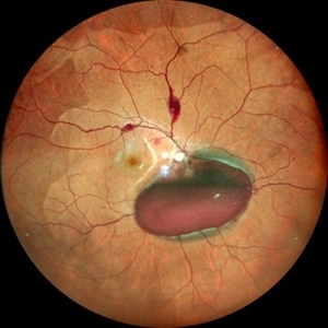
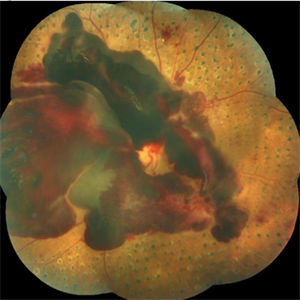
Wide field photograph of 50 year-old woman, known case of JXT in both eyes and known diabetic, after 9 months of PPV for subhyaloid hemorrhage.
Imaging device: Nidek Mirante
Condition/keywords: florid type PDR, JXT, pars plana vitrectomy (PPV)
-
 Valsalva Retinopathy
Dec 20 2021 by Unnati Vishwanath Shukla, M. S. ,DNB, FVRS FNERF, MNAMS,PhD Scholar(Retina)
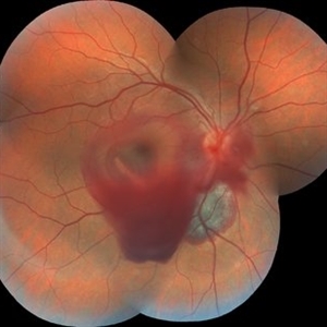
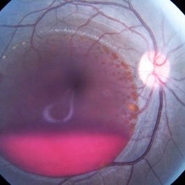
26-year-old male with Valsalva Retinopathy. History of severe cough for 3 days. All hematological investigations were within normal limits.
Photographer: Dr. Unnati Shukla, Consultant, Retina Foundation, Ahmedabad
Imaging device: Nidek Mirante
Condition/keywords: subhyaloid hemorrhage, subretinal hemorrhage, valsalva retinopathy
-
 Subhyaloid Hemorrhage
Mar 1 2021 by Narciso F. Atienza, MD, MBA, FASRS, FPCS, FPAO.
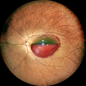
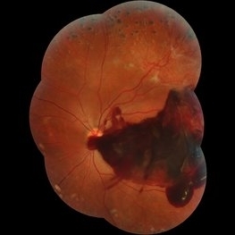
Fundus photo of the left eye, 29-year-old male patient, with previous history of 6 cycles of chemotherapy from Hodgkins lymphoma. Photograph, however looks more like leukemic subhyaloid hemorrhage.
Photographer: Narciso Atienza, Jr. MD, MBA. Legazpi Eye Center
Condition/keywords: Hodgkins lymphoma, subhyaloid hemorrhage
-
 Trio of Retinal Hemorrhages
Dec 8 2020 by Priya Rasipuram Chandrasekaran, MBBS, DO, DNB, FRCS
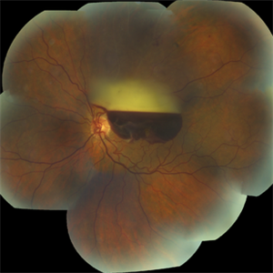
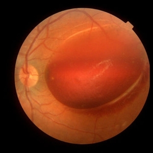
This is the fundus photo of a 29-year-old following blunt trauma showing hemorrhages in all the three layers of the retina (vitreous hemorrhage, subhyaloid hemorrhage and subretinal hemorrhage)
Condition/keywords: blunt trauma, retinal hemorrhage
-
 Subhyaloid Hemorrhage
Jun 10 2020 by Manish Nagpal, MD, FRCS (UK), FASRS
Subhyaloid hemorrhage over macula.
Photographer: Gayathri Mohan, Retina Foundation
Imaging device: NIDEK SLO MIRANTE
Condition/keywords: subhyaloid hemorrhage
-
 Branch Retinal Vein Occlusion with Acute on Chronic Subhyaloid Hemorrhage
Oct 24 2019 by Nichole Lewis
60-year-old male with a branch retinal vein occlusion and subhyaloid hemorrhage and retinal neovascularization. VA HM.
Photographer: Nichole Lewis
Condition/keywords: branch retinal vein occlusion (BRVO), retinal neovascularization, subhyaloid hemorrhage
-
 Proliferative Diabetic Retinopathy (PDR)
Jul 4 2018 by Deepak Bhojwani, MS
Colour Fundus Photograph of a 66-year-old diabetic male with large fibro-vascular proliferative vessels causing subhayolid haemorrhage and tractional retinal detachment involving posterior pole.
Photographer: Deepak Bhojwani
Condition/keywords: diabetes, neovascularization (NV), subhyaloid hemorrhage, tractional retinal detachment
-
 Subhyaloid Hemorrhage, Proliferative Diabetic Retinopathy
May 31 2018 by awaneesh m upadhyay, MBBS, DNB
Right eye fundus photography of a 63-year-old male came with sudden onset defective vision with history of laser photocoagulation done for proliferative diabetic retinopathy.
Photographer: Dr Awaneesh Upadhyay
Condition/keywords: laser photocoagulation, proliferative diabetic retinopathy (PDR), subhyaloid hemorrhage
-
 Valsalva Retinopathy
Apr 6 2018 by Jun Dong Dong
A 45-year-old male with no definite history of activities which can suddenly increase the intrathoracic pressure refused the laser treatment and chose the observation.
Photographer: Jun Dong, Shanghai Aier Eye Hospital
Condition/keywords: subhyaloid hemorrhage
-
 Subhyaloid Hemorrhage
Oct 2 2017 by Mehul A Shah
A 25-year-old patient presented with history of sudden loss of vision following blunt trauma.
Photographer: Mehul Shah
Condition/keywords: subhyaloid hemorrhage
-
 Subhyaloid Hemorrhage With Flat Neovascular Vessels
Sep 26 2017 by Purva Patwari
60-year-old patient with uncontrolled diabetes.
Photographer: Dr Purva Patwari, Patwari Retina Clinic,Ahmedabad, India
Imaging device: Zeiss
Condition/keywords: flat neovascularization
-
 Preretinal Hemorrhage
May 6 2017 by Mitzy E Torres Soriano, MD
Fundus photograph of a 36-year-old-woman with a preretinal subhyaloid hemorrhage (valsalva retinopathy).
Photographer: Mitzy Torres Soriano
Condition/keywords: macular hemorrhage, premacular hemorrhage, preretinal hemorrhage, subhyaloid hemorrhage, valsalva retinopathy
-
 Subhyaloid Hemorrhage
Jul 7 2015 by Hamid Ahmadieh, MD
Color fundus photograph of the left eye of a 25-year-old woman with severe subhyaloid hemorrhage due to an advanced vasoproliferative vitreoretinopathy secondary to a severe idiopathic occlusive retinal vasculitis.
Photographer: Soulmaz Shahmohammad, Negah Eye Center, Tehran, Iran
Condition/keywords: color fundus photograph, occlusive vasculitis, subhyaloid hemorrhage
-
 Subhyaloid Hemorrhage, Right Eye
Jan 28 2015 by Kathy Karsten, COT
Fundus photograph of 38-year-old woman who presented with sudden onset of flashes and a large black floater obscuring vision in the right eye. Recent history of severe coughing and emesis. Diagnosed with subhyaloid hemorrhage and valsalva retinopathy OD.
Photographer: Kathy Karsten, COT
Imaging device: Topcon TRC-50DX
Condition/keywords: subhyaloid hemorrhage
-
 Subhyaloid Hemorrhage
Jun 29 2014 by Woohyok Chang, MD, PhD
Color fundus photograph of the left eye of a 65-year-old man with organizing subhyaloid hemorrhage secondary to branch retinal vein occlusion.
Photographer: Mi-Young Choi, Yeungnam University, Daegu, South Korea
Imaging device: Cannon
Condition/keywords: branch retinal vein occlusion (BRVO), subhyaloid hemorrhage
-
 Primary Subhyaloid Hemorrhage Due to Valsalva Retinopathy
Nov 13 2013 by Hamid Ahmadieh, MD
Color fundus photograph of the left eye of a 25-year-old man with sudden drop of vision due to subhyaloid hemorrhage secondary to Valsalva retinopathy.
Photographer: Soodabeh Fooladin , Negah Eye Center, Tehran
Imaging device: TOPCON OCT
Condition/keywords: subhyaloid hemorrhage, valsalva retinopathy
 Loading…