Search results (16 results)
-
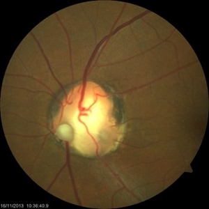
 Venous Loop
Feb 20 2024 by Soobien Lee
A 77-year-old male with a history of bilateral optic neuropathy from bilateral optic nerve sheath meningiomas S/P radiation/proton-beam therapies. Presented with radiation retinopathy OS and a known venous loop OS.
Photographer: Gavin Bragdon, Elman Retina Group
Imaging device: Optos Ultra-Widefield Fluorescein Angiography
Condition/keywords: fluorescein angiogram (FA), Optos, radiation retinopathy, retinal vascular disease, venous loop
-
 Melanocytoma of Optic Disc
Nov 3 2023 by Virginia Gebhart
69 year-old female with pigmented lesion that covers the optic nerve. Patient has been aware for over 30 years. Remains stable and unchanged
Photographer: Virginia Gebhart
Imaging device: Topcon
Condition/keywords: benign melanocytoma, Melanocytoma, optic disc melanocytoma
-
 Optic Nerve Melanocytoma
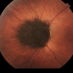
Apr 3 2023 by Gustavo Aguirre Suarez
Fundus photograph of a 36-year-old female with a lesion dependent on the optic nerve head with subretinal extension, elevated, about 1.5 disc diameters, dark brown to black in color, involving more than three quarters of the neuroretinal ring towards the inferonasal area.
Photographer: Dr. Gustavo Aguirre-Suarez
Imaging device: Zeiss Visucam 500
Condition/keywords: melanocytic lesion, Melanocytoma
-
 Gyrate Atrophy
Apr 12 2023 by Ahmed Abbas Hashmi, OD
Left eye fundus of a 53-year-old male patient with advanced gyrate atrophy of the choroid and retina with macular sparing. Optic nerve head is healthy.
Photographer: Ahmed Abbas Hashmi
Imaging device: Topcon TRC-NW8F
Condition/keywords: chorioretinal atrophy
-
 Morning Glory Anomaly
Mar 24 2022 by Elite Bor-Shavit, MD
Disc photo of a 28-years-old male with Morning Glory Anomaly of his right optic nerve observed over time.
Condition/keywords: Morning Glory Anomaly, optic disc
-
 Coloboma involving the Optic nerve, Retina, and Choroid
Dec 6 2021 by Jesus Lozano, MD
78-year-old woman after prophylactic laser photocoagulation (PLP) for her RE Coloboma involving the optic nerve, retina, and choroid. At 6 month follow up, patient preserved her FC vision as it was before the procedure. Retina attached.
Photographer: Yair Bet Yosef, Hadassah Medical Center. Israel
Imaging device: Optos Silverstone fundus image
Condition/keywords: coloboma, coloboma of choroid, coloboma of macula, coloboma of optic disc, PLP, prophylactic photocoagulation
-
 Optic Nerve Avulsion with Vitreous Hemorrhage and Pale Retina
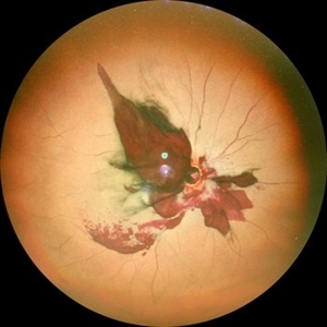
Jan 25 2021 by Sham Talati, DOMS
A 30-year-old male presented with history of trauma to RE with NO Perception of light in the affected eye.
Photographer: Dr. Sham Talati,Retina Foundation,Ahmedabad
Imaging device: Nidek Mirante
Condition/keywords: optic nerve, pale retina
-
 Optic Nerve Pit Right Eye
Feb 15 2021 by Kim Barrett
A 14-year-old male presented with vision loss and VF defect. Patient was treated for presumed amblyopia with patching since age 4. He has had neurologic care for post traumatic skull fracture and brain bleed in 2012. IOP's WNL. OD is without retinoschisis or subretinal fluid. Patient is at risk of serous detachment. Current VA OD 20/200+1 PH 20/80.
Photographer: Kim Barrett C.O.A. Retina Specialist of Michigan, Grand Rapids, MI
Imaging device: Optos California
Condition/keywords: amblyopia, hemifield, Humphrey visual field, nerve, optic nerve pit, visual field defect
-
 Retinoblastoma
Apr 30 2020 by Giselle DeOliveira
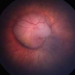
Fundus photograph of an 17-month-old female infant with retinoblastoma over optic nerve.
Photographer: Giselle DeOliveira, University of Miami, Bascom Palmer Eye Institute
Imaging device: Retcam III
Condition/keywords: retinoblastoma
-
 Silicon Droplet in the Cup of Optic Nerve After Multiple Intravitreal Avastin Treatments
Nov 1 2018 by Tammy Mclaughlin
Fundus photograph of a 68-year-old man with silicon droplet in the cup of optic nerve after multiple intravitreal Avastin treatments.
Photographer: Tammy Mclaughlin, Carolina Retina Center, Sumter SC
Imaging device: Zeiss Visucam
Condition/keywords: optic nerve
-
 Post Traumatic Optic Nerve Head Avulsion
Nov 18 2017 by Vishal Agrawal, MD, FRCS,FACS,FASRS
Right eye fundus picture of a 24-year-old male patient who suffered blunt trauma 7 days back with a wooden stick . He presented with NLP vision with a non reacting dilated pupil. Fundus montage picture shows ONH avulsion,CRAO,peripapillary resolving hemorrhages and cicatricial tissue at the edge.
Photographer: Vishal Agrawal, MD, SMS Medical College, Jaipur, India
Imaging device: Zeiss 524
Condition/keywords: avulsion, central retinal artery occlusion (CRAO)
-
 Optic Nerve Head Drusen With Idiopathic CNV
Feb 17 2017 by Kristen Wagner
22-year-old female fundus photograph of a right eye with Optic Nerve Drusen with Idiopathic CNV.
Photographer: Kristen Wagner, COT, OSC Ophthalmic Photographer, Tennessee Retina, Nashville TN
Condition/keywords: choroidal neovascularization (CNV), drusen of optic disc, optic disc drusen
-
 Acute Traumatic Optic Nerve Avulsion
Feb 19 2016 by Mahdi Mwas
Fundus photograph of a 24-year-old gentleman, involved in a road traffic accident resulting in left no perception of light.
Photographer: Mahdi Mwas, FRCS, DRCOphth, Jordan
Condition/keywords: optic nerve head avulsion
-
 Optic Nerve Head Drusen
Feb 12 2015 by Timothy S Fuller, MD
Fundus autofluorescence image of a 34-year-old woman with striking, asymptomatic optic nerve head drusen.
Photographer: Nice Hesse, Texas Retina Associates
Imaging device: Heidelberg Spectralis
Condition/keywords: drusen of optic disc
-
 CMV Retinitis/ Before Treatment
Mar 13 2015 by Niloofar Piri, MD
Fundus photograph of the left eye of a 40-year-old Caucasian female with history of positive HIV test for 23 years. She has been off HAART therapy for the past 2 years and presented with decreased vision OS and upper visual field defect. On examination, she had trace cells in anterior vitreous , hemorrhagic retinitis which starts around the optic nerve and extending to inferotemporal arcade with secondary inferotemporal BRVO; in temporal periphery , she had granular pattern of CMV retinitis which is a manifestation of outer retina involvement.
Photographer: Angela Anderson
Condition/keywords: CMV retinitis, HIV
-
 Optic Nerve Coloboma With 2 Pits, Nasal and Temporal Color
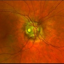
Nov 21 2013 by Alexandre Durao Alves Pereira, MD
Fundus photograph, color, red free, blue lite and FAF of a optic nerve coloboma with 2 pits, one nasal and other temporal.
Photographer: Alexandre Pereira
Imaging device: Visucam 300
Condition/keywords: color photo, optic nerve coloboma
 Loading…