Search results (7 results)
-
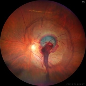 Ruptured Retinal Artery Macroaneurysm
Jun 18 2024 by KANWALJEET HARJOT MADAN, M.S. (Ophthalmology); FAICO (Vitreous - Retina)
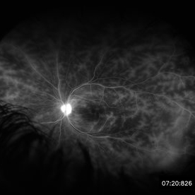
This is a fundus photo depicting ruptured Retinal Artery Macroaneurysm (RAM) in the left eye of a 63 years old female. RAM is an acquired saccular or fusiform dilatation of the retinal arterioles that usually occur within the first three orders of bifurcation. The Superotemporal artery is the most common location. RAM may be asymptomatic or cause a number of complications such as macular edema, serous macular detachment, and hemorrhages.
Photographer: Dr Kanwaljeet Harjot Madan
Condition/keywords: Haemorrhage, macroaneurysm, retinal arteriole
-
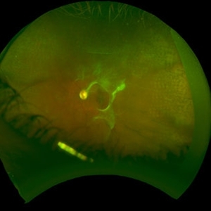 Eales Disease Causing TRD and Macular Edema in Pregnancy
Apr 21 2020 by Richard M Martindale, MD
42-year-old pregnant African American with TRD and peripheral ischemia secondary to Eales disease. She was assigned this diagnosis of exclusion after a thorough work up for other identifiable causes of peripheral ischemia (e.g. sickle cell, syphilis, sarcoid, clotting disorders, SLE, TB, IP, FEVR). We elected to temporize her with PRP and Ozurdex in lieu of anti-VEGF medication given her pregnant status. Note: the Ozurdex pellet is visible in the inferior aspect of this photo.
Photographer: Retina Consultants of Alabama
Imaging device: Optos
Condition/keywords: Eales disease
-
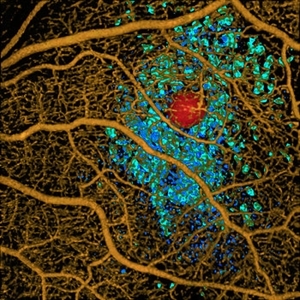 Volume Rendering Structural and Angiographic Optical Coherence Tomography Angiography Image of a Retinal Capillary Microaneurysm, A Newly Described Entity.
May 21 2019 by Richard F. Spaide, MD
This is a newly described entity in which patients develop solitary aneurysms that are much larger than typical microaneurysms and they are supplied by capillaries. The aneurysm is shown in red. The associated macular edema produced cystoid spaces in Henle’s fiber layer, rendered as teal and in the inner nuclear layer as blue.
Photographer: Richard F. Spaide, MD
Condition/keywords: aneurysm, optical coherence tomography (OCT), volume rendering
-
 Retinitis Pigmentosa With Cystoid Macular Edema
Jun 24 2018 by Zachary M Bodnar, MD
64-year-old female with retinitis pigmentosa and cystoid macular edema, both eyes.
Imaging device: optos
Condition/keywords: cystoid macular edema (CME), retinitis pigmentosa
-
 Retinal Vasculitis in Behcet's OS
Jun 29 2018 by Gareth Lema, MD, PhD
IVFA at 7 minutes showing retinal vasculitis, cystoid macular edema, and disc staining.
Photographer: Ross Eye Institute, University at Buffalo Jacobs School of Medicine, Buffalo. NY
Imaging device: Optos
Condition/keywords: Behcet's Disease, cystoid macular edema (CME), disc staining, retinal vasculitis
-
 Multiple Myeloma with Cytomegalovirus Retinitis
Apr 5 2018 by Kim Barrett
Ultra-wide field fluorescein angiogram of a 77-year-old male with multiple myeloma. Patient's angiogram presented significant peripheral retinal ischemia and cystoid macular edema. Patient tested positive for polymerase chain reaction, confirming cytomegalovirus retinitis. Patient is being treated with intravitreal ganciclovir and his current vision is 20/200.
Photographer: Kim Barrett, COA
Imaging device: Optos
Condition/keywords: cystoid macular edema (CME), fluorescein angiogram (FA), fluorescein leakage, intravitreal ganciclovir, myeloma, peripheral ischemia, positive polymerase chain reaction (PCR), ultra-wide field imaging
-
 SLE Retinopathy
Nov 14 2016 by Mitzy E Torres Soriano, MD
25-year-old female patient with systemic lupus erythematosus. Photographs show cotton wool spots, intraretinal hemorrhages and vascular tortuosity. FA demonstrated retinal vasculitis and OCT revealed cystoid macular edema. In this case diagnosis of SLE was made after ocular manifestation.
Photographer: Grupo Laser Vision, Rosario, Argentina
Condition/keywords: cotton wool spots, occlusive retinal vasculitis, occlusive vasculitis, systemic lupus erythematosus, vasculopathy
 Loading…