Search results (33 results)
-
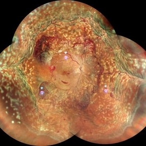
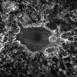
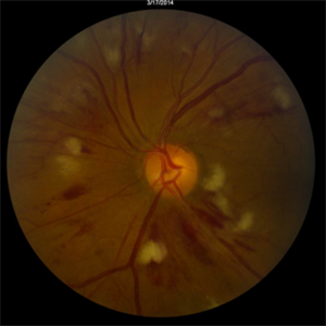
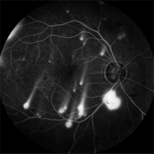 Shooting Stars
Jul 9 2025 by Majda Hadziahmetovic, MD
Fluorescein angiography image demonstrating multiple areas of neovascularization in a middle-aged male patient with long-standing diabetes.
Condition/keywords: proliferative diabetic retinopathy (PDR)
-
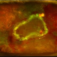
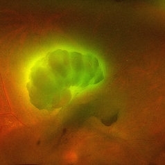 Aurora Borealis in Retina
Apr 25 2025 by Poornachandra B, MS, FVRS
Fundus picture of 54 year old male with proliferative diabetic retinopathy with fluorescent blood clot in vitreous cavity.
Photographer: Mr Dhikshith
Imaging device: Optos daytona
Condition/keywords: blood, proliferative diabetic retinopathy (PDR)
-
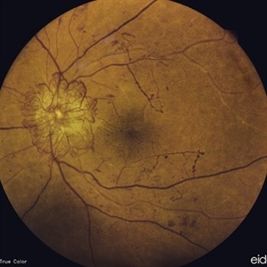
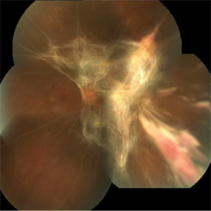
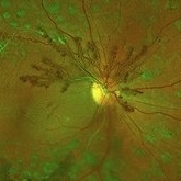 Proliferative Diabetic Retinopathy
May 2 2024 by Aditya S Kelkar, MS, FRCS, FASRS,FRCOphth
This fundus photo captures an intricate web of new vessels at optic disc.
Photographer: Dr Yash Garg , National Institute of Ophthalmology , Pune
Imaging device: OPTOS DAYTONA
Condition/keywords: web of collaterals
-
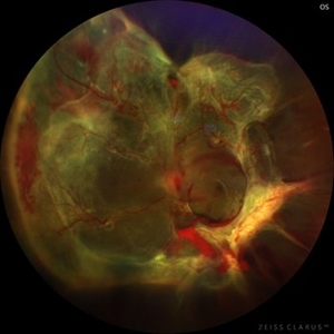
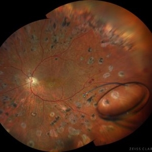 Intraocular lens luxated to the vitreous cavity
Jun 24 2023 by Mariam Cernichiaro-Espinosa, MD
Three-piece intraocular lens luxated to the vitreous cavity in a patient with photocoagulated diabetic retinopathy after blunt trauma.
Photographer: Mariam Cernichiaro-Espinosa, Asociación para Evitar la Ceguera en México, I.A.P. Mexico City, Mexico.
Imaging device: Zeiss Clarus
Condition/keywords: diabetic retinopathy, intraocular lense in vitreous, lens luxation
-
 Candy Stripe Sign
Mar 30 2023 by pedro fernandes souza neto
Candy Stripe Sign, patient with proliferative diabetic retinopathy progressing to vitreous hemorrhage and subsequently to ghost cell glaucoma.
Photographer: Marlos Henrique Oliveira Junior, Federal University of Bahia.
Condition/keywords: dehemoglobinized hemorrhage, diabetes, diabetic glaucoma
-
 Diabetic traction retinal detachment
Jan 9 2023 by JORGE SOBERANES
Proliferative diabetic retinopathy with extensive traction retinal detachment in a patient with type 1 diabetes mellitus.
Photographer: Dr. Jorge I. Soberanes, Asociación para Evitar la Ceguera en México.
Imaging device: Zeiss Clarus 700
Condition/keywords: Retinal Detachment, tractional retinal detachment
-
 High risk Proliferative Diabetic Retinopathy treated with Pan Retinal Photocoagulation
Nov 5 2022 by Somnath Chakraborty, MD
A Fundus Photo Montage of 43 year old Asian Male with Type 2 Diabetes Mellitus since 7 years who presented with sudden onset diminition of vision in his Left eye. BCVA OS was 20/200. He was diagnosed to have Pre retinal bleed due to Proliferative Diabetic Retinopathy and was treated with Pan Retinal Photocoagulation. This image shows a large neo-cascular frond at the disc and superior to it with Pre-retinal bleed and Fresh laser marks along
Photographer: Pulak Roy
Condition/keywords: diabetic blindness, diabetic retinopathy vitrectomy study (DRVS), fresh laser burns, laser photocoagulation, preretinal hemorrhage, proliferative diabetic retinopathy (PDR)
-
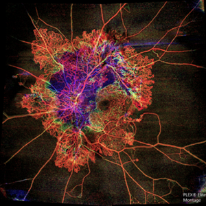
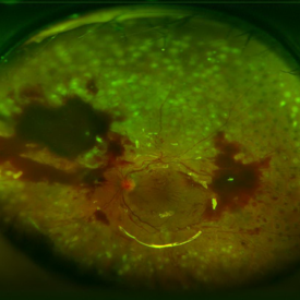
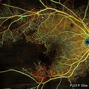 Proliferative Diabetic Retinopathy with Macular Isquemia by OCT Angiography
Oct 14 2022 by JORGE SOBERANES
Depth-encoded OCT angiography of a patient with proliferative diabetic retinopathy showing vascular changes and extensive ischemia including macular area.
Photographer: Jorge I. Soberanes, Asociación para Evitar la Ceguera en México.
Imaging device: PLEX Elite 9000, Zeiss
Condition/keywords: diabetic retinopathy, OCT Angiography
-
 Proliferative Diabetic Retinopathy
Jul 15 2022 by Gabriel Costa Andrade, PhD
Fundus angiography of an 22-year-old man with proliferative diabetic retinopathy and macular ischemia.
Photographer: Dr Gabriel Andrade
Condition/keywords: Diabetes
-
 JXT and Proliferative Diabetic Retinopathy
Jan 13 2022 by ASRS Staff
Wide field photograph of 50 year-old woman, known case of JXT in both eyes and known diabetic, after 9 months of PPV for subhyaloid hemorrhage.
Imaging device: Nidek Mirante
Condition/keywords: florid type PDR, JXT, pars plana vitrectomy (PPV)
-
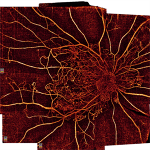 Proliferative Diabetic Retinopathy
Oct 16 2021 by Timur Shaimov
32 y.o. female with Type 1 Diabetes with no glucose compensation for several years. A manual montage of several 8x8 mm OCT angiograms were obtained for this Widefield OCTA image.
Photographer: Timur Shaimov
Imaging device: RTVue xR Avanti
Condition/keywords: OCT Angiography, proliferative diabetic retinopathy (PDR)
-
 Venous Beading
Nov 4 2021 by Stefanie Palmer
Venous Beading in a patient with both PDR and CRVO.
Photographer: Stefanie Palmer, CRA
Imaging device: Topcon
Condition/keywords: central retinal vein occlusion (CRVO), diabetic retinopathy, proliferative diabetic retinopathy (PDR), venous beading
-
 Diabetic Retinopathy Treated with PRP Laser
Jun 8 2021 by Ronald Coriasso
Diabetic retina treated with complete 360 PRP laser, taken during fluorescein angiogram.
Photographer: Ronald Coriasso
Imaging device: OPTOS
Condition/keywords: pan-retinal photocoagulation (PRP)
-
 Proliferative Diabetic Retinopathy with Choroidal Effusion Status Post PRP
Dec 15 2020 by Manish Nagpal, MD, FRCS (UK), FASRS
A 17-year-old juvenile diabetic patient came to us with extensive neovascular proliferations and PRP done a week back and had developed 360 degree choroidal effusion as seen in this wide field montage image
Photographer: Sham Talati, Retina Fellow , Retina Foundation, Ahmedabad, India
Imaging device: Mirante CSLO
Condition/keywords: choroidal effusion, diabetic retinopathy, proliferative diabetic retinopathy (PDR)
-
 Proliferative Diabetic Retinopathy with Traction Retinal Detachment OS
Apr 28 2020 by Pauline T Merrill, MD, FASRS
Fundus photographs of an 29-year-old man with PDR, TRD, VH OS.
Photographer: Karen Parque, Illinois Retina Associates
Condition/keywords: proliferative diabetic retinopathy (PDR), tractional retinal detachment
-
 Diabetic Macular TRD
Jan 10 2020 by Somnath Chakraborty, MD
Fundus Montage image of the left eye of a 48-year-old type 2 diabetic with post PRP macular extensive tractional retinal detachment involving macula.
Photographer: Pulak Roy
Condition/keywords: diabetic retinopathy, proliferative diabetic retinopathy (PDR), tractional retinal detachment, vitrectomy, vitreomacular surgery
-
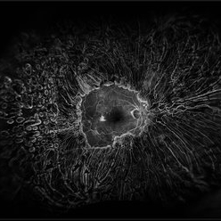
 Flame of the Forest
Apr 9 2020 by Daraius N Shroff, MS FMRF FRCS
A 54-year-old man with DM for 15 years. The left eye had a visual acuity of 20/40. Wide field swept source OCTA revealed branching out central neovascular trunk vessels from the disc with terminal loops, along with exuberant proliferation of irregular small-calibre fine new vessels. The patient underwent OCTA guided pan retinal photocoagulation.
Photographer: Anuj Choudhary, Shroff Eye Centre, New Delhi
Imaging device: Zeiss Plex Elite 9000
Condition/keywords: proliferative diabetic retinopathy (PDR)
-
 Retinal Detachment with PVR (s/ SPR, PPV, MPV, 360 Retinectomy, PFO, PI, FAx, SO)
Aug 22 2019 by Merrick Avila
Ultra-wide field pseudocolor fundus photograph of a 64-year-old female with a treated retinal detachment with proliferative vitreoretinopathy. Patient has a history of complex retinal detachments that have been treated multiple times. On exam 8-22-19, there were large macular holes with LP vision. There was a long discussion about guarded nature of her condition and goals or trial for repair including globe sparing prevention of phthisis.
Photographer: Merrick Avila
Imaging device: Optos
Condition/keywords: diabetic retinopathy, hemorrhage, Optos, proliferative vitreoretinopathy (PVR), retinectomy, silicone oil
-
 Proliferative Diabetic Retinopathy
Jul 9 2019 by Chinmayi Vyas
38-year-old type 1 diabetic female with gross neovascularization of disc.
Photographer: Sangeeta
Condition/keywords: neovascularization of the disc (NVD)
-
 PDR; High Myopia; PRP
May 2 2019 by Carissa Hurdstrom
PDR; high myopia; PRP
Imaging device: Optos
Condition/keywords: fluorescein angiogram (FA), high myopia, pan-retinal photocoagulation (PRP), proliferative diabetic retinopathy (PDR)
-
 Proliferative Diabetic Retinopathy (PDR)
Jul 4 2018 by Deepak Bhojwani, MS
Colour Fundus Photograph of a 66-year-old diabetic male with large fibro-vascular proliferative vessels causing subhayolid haemorrhage and tractional retinal detachment involving posterior pole.
Photographer: Deepak Bhojwani
Condition/keywords: diabetes, neovascularization (NV), subhyaloid hemorrhage, tractional retinal detachment
-
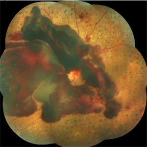
 Extensive Tractional Retinal Detachment in Proliferative Diabetic Retinopathy
Jun 4 2018 by Diva Kant Misra, MBBS, DO, DNB, MNAMS, FVRS
Montage fundus photograph of a 54-year-old male diabetic patient showing extensive TRD with PDR.
Photographer: DIVA KANT MISRA
Condition/keywords: diabetes, proliferative diabetic retinopathy (PDR), tractional retinal detachment
-
 Subhyaloid Hemorrhage, Proliferative Diabetic Retinopathy
May 31 2018 by awaneesh m upadhyay, MBBS, DNB
Right eye fundus photography of a 63-year-old male came with sudden onset defective vision with history of laser photocoagulation done for proliferative diabetic retinopathy.
Photographer: Dr Awaneesh Upadhyay
Condition/keywords: laser photocoagulation, proliferative diabetic retinopathy (PDR), subhyaloid hemorrhage
-
 TRD After Surgical Repair
Sep 14 2017 by Theodore Leng, MD, MS, FASRS
TRD after surgical repair.
Condition/keywords: proliferative diabetic retinopathy (PDR), tractional retinal detachment
-
 Radiation Retinopathy
Mar 12 2016 by David Callanan, MD
55-year-old with background diabetic retinopathy that developed renal cell carcinoma. Underwent radiation to left orbit.
Condition/keywords: radiation maculopathy
 Loading…