Search results (119 results)
-
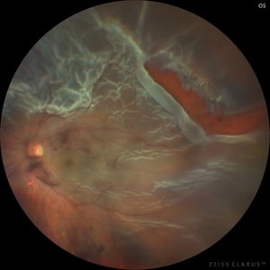
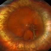 Repaired Retinal Detachment with Scleral Buckle
Mar 25 2025 by Kimberly Wakester
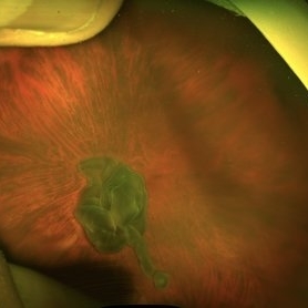
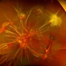
Optomap RGB montage of an 64-year-old woman with a repaired retinal detachment with scleral buckle in the right eye. There is nasal and inferior pre-retinal membranes with traction. PPV was recommended but patient defers to proceed with sx at this time. Will continue to follow patient closely for worsening traction. Patient was educated on how to monitor their peripheral vision and was advised to report any changes immediately.
Photographer: Kimberly Wakester, COA, OCT-C
Imaging device: Optos California
Condition/keywords: pre-retinal membrane with traction, repaired RD, scleral buckle
-
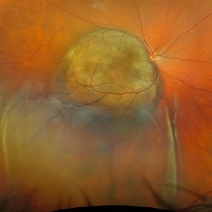
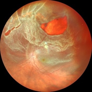
 A Large Break at the Posterior Pole With RD With PVR (S/p Old Blunt Trauma)
Jan 16 2025 by Anand Temkar
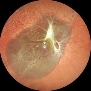
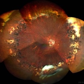
Right eye widefield fundus color photo of a 10 year old kid who noticed diminution of vision in right eye since a month. We can see the large break at the posterior pole with rolled up margins associated with retinal detachment and PVR changes.
Photographer: Dr.Anand Temkar- Retina Foundation, Ahmedabad
Imaging device: Mirante
Condition/keywords: posterior pole break, proliferative vitreoretinopathy (PVR), Retinal Detachment
-
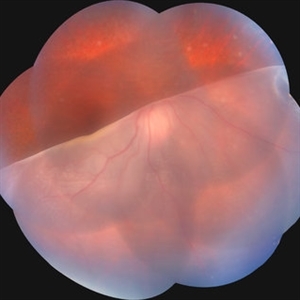
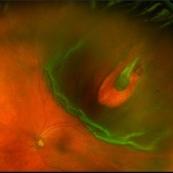
 Hemorrhagic Choroidals
Jan 22 2025 by Danish Shabbir, Ophthalmic Technologist
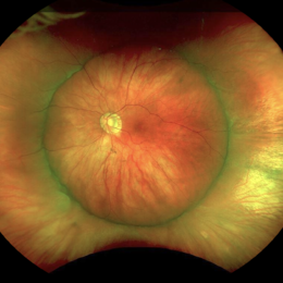
78 year old female complains of suddenly vision decrease 2 days ago.
Photographer: Danish Shabbir,Retina-EyeCare Centre
Imaging device: Optos California
Condition/keywords: choroidal detachment, Retinal Detachment, retinal detachment with choroidal
-
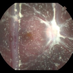
 Choroidal Melanoma with Serous Retinal Detachment
Dec 20 2024 by Daniel Davis, OCT-C
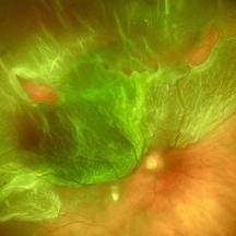
67 year old male presenting with large pigmented choroidal mass with serous retinal detachment.
Photographer: Daniel Davis, OCT-C, The Retina Institute
Imaging device: Optos California
Condition/keywords: Retina detachment
-
 Giant Retinal Tear
Jul 15 2024 by Arthi Mohankumar , MS,MRCS ED, FICO,FAICO
Fundus montage of a 15 year old boy with Marfans syndrome who presented with defective vision in the right eye.
Photographer: Arthi Mohankumar
Condition/keywords: giant retinal tear, Retinal detachment
-
 Fish Hook Eye Trauma
Jun 12 2024 by Miguel Brito, MD, FASRS
Fundus photograph of a 15-year-old boy post cataract aspiration, pars plana vitrectomy, suprachoroidal drainage, and retinal reattachment surgery secondary to traumatic endophthalmitis.
Photographer: Miguel Brito
Condition/keywords: endophthalmitis, PFCL, Retinal detachment under Silicon Oil, retinal fold
-
 Post Combined Surgery of Cataract, TRD & Vitreous Hemorrhage
Jun 27 2024 by Sanauddin Samejo , Diploma (Ophthalmic Technician Training Course)
A 27 year-old diabetic female visited the clinic one week after combined surgery of cataract, tractional retinal detachment and vitreous hemorrhage.
Photographer: Sanauddin Samejo, Burjeel Hospital, Abu Dhabi, UAE
Imaging device: Silver Stone Optos
Condition/keywords: Combined Surgery Cataract Tractional Retinal Detachment Vitreous Hemorrhage, POST SURGERY, Retinal Detachment, TRD
-
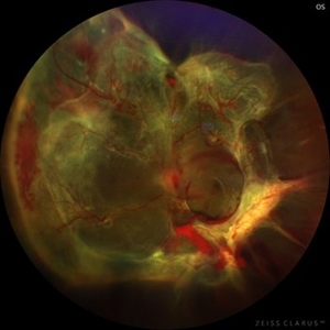
 Fundus Photo of Closed Funnel Retinal Detachment
Apr 10 2024 by Max D Schlesinger, MD
Wide-field funds photography of a closed funnel retinal detachment; patient had previously undergone 360 degree retinectomy in attempt to re-attach retina for a chronic retinal detachment, which was unsuccessful.
Condition/keywords: Closed funnel RD, detachment, Optos
-
 The Bullet Ridden Retina
Feb 17 2024 by SHISHIR VERGHESE, MS, FVRS, FAICO (Retina)
Fundus image obtained of a case of lasered branch retinal vein occlusion (BRVO) with fibrovascular proliferation (FVP) where the laser marks have given way to multiple small retinal holes due to traction from the same.
Photographer: DIVYA SHAJI
Imaging device: NIDEK MIRANTE
Condition/keywords: BRVO, chronic retinal detachment
-
 Post-Operative Scleral Buckle
Mar 8 2024 by Ethan K Sobol, MD
The post operative week one appearance of a macula-on retinal detachment repaired with a 5mm strip encircling band, cryotherapy, and external drainage.
Photographer: Bryan Murphy, Senior Ophthalmic Photographer (Retina Group of Washington)
Imaging device: Optos California
Condition/keywords: scleral buckle
-
 New Retinal Detachment 6w s/p RD repair
Nov 16 2023 by Virginia Gebhart
13 year old male presented with new blind spot 6 weeks s/p RD repair with cryo/scleral buckle/prophylaxis laser with gas bubble. New RD involving the macula, posterior to scleral buckle, secondary to PVD. Small gas bubble remaining. Pt was brought back to OR for repeat PPV and silicone oil repair
Photographer: Virginia Gebhart
Imaging device: Optos
Condition/keywords: gas bubble, Retinal Detachment, retinal detachment of the macula, scleral buckle
-
 Rhegmatogenous Retinal Detachment
Sep 4 2023 by Kayne Michael McCarthy, MD, MPH
Fundus photograph of a 59-year-old man with a rhegmatogenous retinal detachment and multiple retinal tears.
Photographer: Gaurav Shah MD, West Coast Retina, San Francisco
Imaging device: Optos p200dtx
Condition/keywords: Retinal Detachment, rhegmatogenous retinal detachment, tears
-
 Total Rhegmatogenous retinal detachment with lattice degeneration & Vitreous haemorrhage
Jul 31 2023 by Harsh Vardhan Singh, MS
72-year male presented PVD induced total retinal detachment with vitreous hemorrhage
Photographer: Dr Harsh Vardhan Singh, AIIMS, Guwahati
Imaging device: Zeiss Clarus 700
Condition/keywords: chronic retinal detachment, hemorrhage, rrd
-
 Retina Detachment
May 1 2023 by RAKESH SHAH, MS DNB FACS FRF FICO MBA
39 year-old female came with sudden loss of vision, on examination rhegmatogenous retinal detachment with large horse shoe tear and linear tear noted
Photographer: Dr.Rakesh shah
Imaging device: Nidek Mirante machine
Condition/keywords: rhegmatogenous retinal detachment
-
 Macula on Retinal Detachment with large Horseshoe Tear
Apr 26 2023 by Kelli Nyenhuis
Optos photograph of a 61-year-old male with a macula on retinal detachment and large horseshoe tear. Patient had no visual changes.
Photographer: Kelli Nyenhuis, COA
Imaging device: Optos California
-
 Retinal detachment
Apr 12 2023 by Ahmed Abbas Hashmi, OD
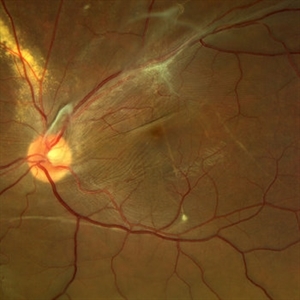
Color fundus photograph of the left eye of a 30-year-old man with asymptomatic inferior retinal detachment with pigmented demarcation line. Macula and Disc healthy.
Photographer: Ahmed Abbas Hashmi
Imaging device: Topcon TRC-NW8F
Condition/keywords: Pigmentary demarcation line, Retinal Detachment
-
 Macula Off Retinal Detachment
Mar 22 2023 by Zach Seim
An ultra-widefield fundus image of a 65 year old male with a Macula Off Retinal Detachment. Patient's vision at the time of the image was CF at 6 Feet and surgical options were discussed. Fluid-gas exchange was performed without complications.
Photographer: Zach Seim
Imaging device: Optos California
Condition/keywords: left eye, macula off retinal detachment, OPTOS CALIFORNIA, scanning laser ophthalmoscope, ultra-widefield image
-
 Rhegmatogenous Retinal Detachment
Feb 26 2023 by Aditya S Kelkar, MS, FRCS, FASRS,FRCOphth
Color fundus photograph of left eye showing rhegmatogenous retinal detachment.
Photographer: Dr. Sahil Wagh, National Institute of Ophthalmology, Pune, India.
Imaging device: Zeiss Clarus 500
Condition/keywords: Retinal Detachment, rhegmatogenous retinal detachment
-
 Diabetic traction retinal detachment
Jan 9 2023 by JORGE SOBERANES
Proliferative diabetic retinopathy with extensive traction retinal detachment in a patient with type 1 diabetes mellitus.
Photographer: Dr. Jorge I. Soberanes, Asociación para Evitar la Ceguera en México.
Imaging device: Zeiss Clarus 700
Condition/keywords: Retinal Detachment, tractional retinal detachment
-
 Choroidal Melanoma with Exudative Retinal Detachment
Mar 2 2023 by Aditya S Kelkar, MS, FRCS, FASRS,FRCOphth
Color fundus photograph of the left eye of a 45 year old male showing choroidal melanoma with exudative retinal detachment.
Photographer: Dr. Pranali Surawase, National Institute of Ophthalmology, Pune, India.
Imaging device: Zeiss Clarus 500
Condition/keywords: choroidal mass, exudative retinal detachment, Retinal detachment
-
 Choroidal Melanoma
Nov 3 2022 by pedro fernandes souza neto
Transillumination of Enucleation specimen of Choroidal Melanoma: anterior chamber is closed. Total secondary retinal detachment with subretinal serous fluid and some subretinal hemorrhages are present.
Photographer: Eduardo Marback, Federal University of Bahia, Brazil
Condition/keywords: enucleation, melanoma
-
 Posteriorly dislocated IOL
Oct 22 2022 by Vishal Agrawal, MD, FRCS,FACS,FASRS
67 yr old male , post PPV for retinal detachment ( 5 years ) presented with sudden DOV . On examination posteriorly dislocated 4 loop haptic iol - bag complex was noted .
Photographer: Pankaj
Imaging device: CLARUS 700
Condition/keywords: dropped intraocular lens (IOL)
-
 Total retinal Detachment multiple holes
Sep 26 2022 by Denica Rodriguez
60 year old Male presented with two week old Macula off Retinal detachment with multiple tears.
Photographer: Denica Rodriguez
Imaging device: Optos California
Condition/keywords: color fundus photograph, color photo, macula-off, optos, pseudocolor, Retinal detachment, retinal holes, retinal tear, Retinal tear with detachment, superior arcade, superior field, superior retina, total retinal detachment
-
 Displaced & folded macula
Oct 10 2022 by Ricardo Leitão Guerra
Tractional retinal detachment due to sickle cell retinopathy leading to a displaced and folded appearance of the macula in this 36-yo male. Subretinal bands are also noticed crossing the macula towards inferior retinal detachment area.
Photographer: Ricardo Leitão Guerra
Imaging device: Clarus 700 - Zeiss
Condition/keywords: folds, sickle cell retinopathy, subretinal bands, tractional retinal detachment
-
 Sickle Cell Retinopathy
Nov 5 2022 by Mateus Queiroz Corrêa, MD
19 -year-old young man with combined rhegmatogenous and tractional retinal detachment secondary to a proliferative sickle retinopathy ( stage V)
Photographer: Mateus Corrêa, Sorocaba Eye Bank Hospital
Imaging device: Optos California
Condition/keywords: Retinal detachment, sickle cell retinopathy
 Loading…