Search results (17 results)
-
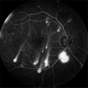 Shooting Stars
Jul 9 2025 by Majda Hadziahmetovic, MD
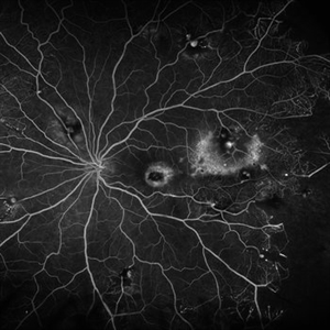
Fluorescein angiography image demonstrating multiple areas of neovascularization in a middle-aged male patient with long-standing diabetes.
Condition/keywords: proliferative diabetic retinopathy (PDR)
-
 Venous Loop
Feb 20 2024 by Soobien Lee
A 77-year-old male with a history of bilateral optic neuropathy from bilateral optic nerve sheath meningiomas S/P radiation/proton-beam therapies. Presented with radiation retinopathy OS and a known venous loop OS.
Photographer: Gavin Bragdon, Elman Retina Group
Imaging device: Optos Ultra-Widefield Fluorescein Angiography
Condition/keywords: fluorescein angiogram (FA), Optos, radiation retinopathy, retinal vascular disease, venous loop
-
 Lady in a dress
Feb 9 2023 by Shelby Helton
Fluorescein Angiography on a 67-year-old male with significant RPE changes secondary to a severe subretinal hemorrhage that required a vitrectomy with subretinal TPA in 2013.
Photographer: Shelby Helton
Imaging device: Heidelberg Spectralis
Condition/keywords: wet age-related macular degeneration (wet AMD)
-
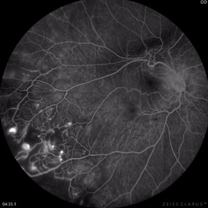 Coats Disease Fluorescein Angiography
Sep 2 2022 by FLOR ANGELICA JACOME GUTIERREZ
Fluorescein angiography of a patient with Coats disease where we found telangiectatic vessels, aneurysms, peripheral capillary nonperfusion and perivascular leak.
Photographer: Dr.Guillermo Salcedo Villanueva
Imaging device: Zeiss CLARUS 700 (FA)
Condition/keywords: Coats' disease, epiretinal membrane (ERM)
-
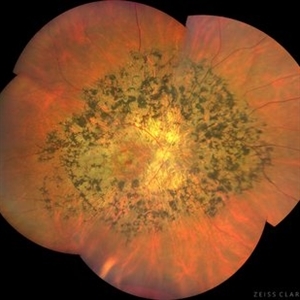 Tapetoretinal Degeneration
Sep 7 2022 by JEFFERSON R SOUSA, Tecg.º (Biomedical Systems Technology)
Patient 52 years old, Male, progressive loss of vision since the age of 20. Retinography showed mobilization of pigments in osteoblasts, extensive area of atrophy of the pigmentary epithelium and choroid. On fluorescein angiography, typical changes following the characteristic patterns of paracentra retinal retinitis pigmentosa. Autofluorescent fundus with a sectorial autohypofluorescence pattern in the regions of atrophies.
Photographer: JEFFERSON ROCHA DE SOUSA - Retinal Department at Instituto Dr. Suel Abujamra Sao Paulo-Brazil
Imaging device: Clarus 700 - Zeiss, composite of four 135 degree images.
Condition/keywords: pericentral retinitis pigmentosa, tapeoretinal degeneration
-
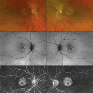 CERKL-related Cone Rod Dystrophy
Jun 27 2022 by Hanna Choi
37-year-old female with cone-rod dystrophy. Developed photophobia and progressive blurry vision in the third decade. VA 20/40 OD, 20/30 OS. The patient is compound heterozygous for pathogenic mutations in the CERKL gene (Arg465Trp and Arg283*).
Photographer: Kaitlynn Silva, New England Retina Consultants
Imaging device: Ultrawide-field Optos Fundus Photography, Autofluorescence, Fluorescein Angiography
Condition/keywords: cone dystrophy, inherited retinal disease, maculopathy
-
 Idiopathic retinal vasculitis, aneurysms and neuroretinitis
Apr 24 2022 by Aniruddha K Agarwal, MD
Ultra-wide field fundus fluorescein angiography (FFA) of the left eye from an asymptomatic, healthy 33-year-old woman who was referred to the retina clinic from a refractive surgery unit due to the presence of vascular anomalies and hard exudates in both eyes. FFA revealed the characteristic sacular aneurysms at the bifurcation of retinal arterioles in the posterior pole, together with microvascular anomalies and capillary closure peripherally.
Photographer: Julio J GONZALEZ-LOPEZ, MD, PhD, FEBO and Teresa GONZALEZ-LOMAS, RN
Imaging device: Optos California
Condition/keywords: IRVAN Syndrome, IUSG, neuroretinitis, retinal vasculitis, uveitis
-
 Coats Disease
Feb 18 2022 by Ahmad B. Tarabishy, MD
43 year old gentleman with poor vision in his left eye for many years. Examination shows multiple retinal telangiectasias and aneurysms. Ultrawide field fluorescein angiography shows light-bulb aneurysms, telangiectasias, and extensive vascular remodeling and non-perfusion.
Photographer: Dr. Angela Rico, Retina Specialists of Tampa
Condition/keywords: Coats' disease
-
 Coats' Disease
Feb 2 2021 by Niloofar Piri, MD
#2 Fluorescein angiography of the same patient in lamellar arteriovenous phase, demonstrating temporal peripheral telangiectatic vessels, as well as hyperfluorescent aneurysma lesions. Note the anterior capillary non perfusion. Posterior hypofluorescence is secondary to blocking effect from hard exudates.
Condition/keywords: Coats' disease, Leber's miliary aneurysm
-
 Lupus Retinopathy
Mar 14 2021 by Marco Antonio Sauza
Fluorescein angiography photo of and 13-year-old female with ischemic retinopathy with LES.
Photographer: Marco Sauza
Imaging device: Zeiss fundus camera
Condition/keywords: systemic lupus erythematosus (SLE) retinopathy
-
 Wyburn-Mason Fluorescein Angiography
Apr 29 2018 by Sarina M Amin, MD
Wide-field fluorescein angiography of a 32-year-old woman with Wyburn-Mason syndrome showing temporal periphery capillary nonperfusion.
Photographer: Sarah Ellano, Retinal Consultants of Arizona, Phoenix, Arizona
Imaging device: Optos
Condition/keywords: Wyburn-Mason
-
 Retinitis Pigmentosa With Hemangioma CF
Dec 15 2016 by Manish Nagpal, MD, FRCS (UK), FASRS
Fluorescein angiography OS of a patient having retinitis pigmentosa with a hemangioma inferiorly.
Condition/keywords: hemangioma, retinitis pigmentosa
-
 Retinal Dystrophy of 24-Year-Old Male/ AF OD
Nov 25 2015 by Zach Dupureur
Fluorescein angiography of a 24-year-old male. Juvenile retinoschisis on OCT. FA shows outer retinal staining. Could be associated with Goldman Farve Syndrome.
Photographer: Zach Dupureur OCT-C
Imaging device: Heidelberg Spectralis
Condition/keywords: Goldmann-Favre Syndrome, juvenile retinoschisis, retinal dystrophy
-
 Ischemic Branch Retinal Vein Occlusion With Compensatory Collateral Vessels
Jul 8 2015 by Kathy Karsten, COT
Heidelberg fluorescein angiography picture of ischemic branch retinal vein occlusion with compensatory collateral vessels in 30-year-old woman.
Photographer: Kathy Karsten, COT
Imaging device: Heidelberg capturing system
Condition/keywords: ischemia
-
 Sea Fan Neovascularisation
Apr 27 2015 by Neha Goel, MS DNB FRCS (Glasg)
Fluorescein angiography of the left eye of a 40-year-old male.
Photographer: Neha Goel
Imaging device: Zeiss visucam
Condition/keywords: Eales disease, neovascularization elsewhere (NVE), vasculitis
-
 Chronic Central Serous Chorioretinopathy (CSCR)
Nov 15 2014 by Rita Couceiro, MD, MS
53-year-old black male, with no relevant prior medical history, complained of bilateral blurry vision for the previous 16 years. On examination, visual acuity was 20/50 on the right eye (OD) and 20/100 on the left eye (OS). Anterior segment evaluation was unremarkable. Fundoscopy revealed pigmentary changes near the macular area in both eyes, with a mottling configuration, suggesting chronic CSCR. Fluorescein angiography showed an ink-blot pattern, with leakage superior to the fovea in OD and nasal to the fovea in OS.
Photographer: Telma Gala - Hospital de Santa Maria, Lisbon, Portugal
Condition/keywords: chronic central serous chorioretinopathy (CSCR)
-
 Aggressive Posterior Retinopathy of Prematurity (AP-ROP)
Nov 21 2013 by Rini Hersetyati, MD
This child with AP-ROP was born 31 weeks gestational age and 1700g birth weight. He was imaged at 38 weeks.
Photographer: Dr. Rini Hersetyati, Klinik Mata Nusantara, Jakarta, Indonesia
Imaging device: RetCam Fluorescein Angiography
Condition/keywords: aggressive posterior retinopathy of prematurity (APROP)
 Loading…