File number: 28152
Comments
-
 Sarina M Amin, MD (June 7 2018)
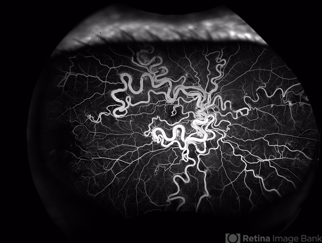
Thank you for your comment! Visual acuity in the eye shown is 20/40; the fellow eye is 20/20 with normal appearing fundus. There is some early nonperfusion in the temporal periphery of the affected eye which we are monitoring. Neuroimaging was also normal.
-
 Suber S. Huang, MD, MBA, FASRS (June 7 2018)
Super capture. Vision and status of fellow eye would be useful. Thanks for sharing!
Sign in to comment.
 Initializing download.
-
 By Sarina M Amin, MD
Co-author(s): Sarina Amin and Neal Palejwala, Retinal Consultants of Arizona, Phoenix, Arizona - Uploaded on Apr 29, 2018.
- Last modified by Caroline Bozell on Jul 13, 2018.
- Image of the week
-
Jul 15, 2018
View all images of the week - Rating
- Appears in
- Miscellaneous
- Condition/keywords
- Wyburn-Mason
- Photographer
- Sarah Ellano, Retinal Consultants of Arizona, Phoenix, Arizona
- Imaging device
-
Fundus camera
Optos - Description
- Wide-field fluorescein angiography of a 32-year-old woman with Wyburn-Mason syndrome showing temporal periphery capillary nonperfusion.


")
")
")


