Search results (282 results)
-
 Active CNVM
Jul 12 2023 by Harsh Vardhan Singh, MS
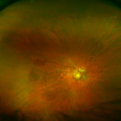
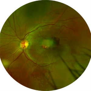
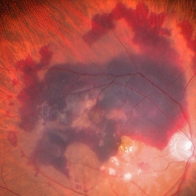
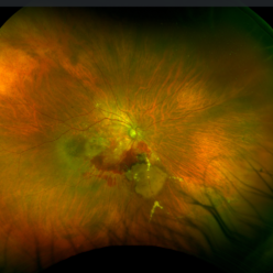
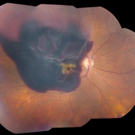
55-year male with left eye sub-retinal hemorrhage due to Active CNVM, Colour fundus photograph of left eye subretinal hemorrhage due to Active CNVM
Photographer: Harsh Vardhan Singh
Condition/keywords: choroidal neovascular membrane (CNVM), CNVM, subretinal hemorrhage
-
 Active CNVM
Jul 12 2023 by Harsh Vardhan Singh, MS
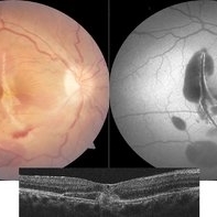
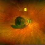
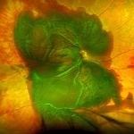
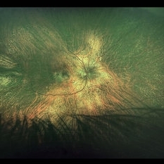
55-year male with left eye sub-retinal hemorrhage due to Active CNVM, Colour fundus photograph of left eye subretinal hemorrhage due to Active CNVM; Red-free image of left eye sub-retinal hemorrhage due to Active CNVM
Photographer: Harsh Vardhan Singh
Condition/keywords: choroidal neovascular membrane (CNVM), CNVM, subretinal hemorrhage
-
 ARMD, SRNVM
Aug 7 2013 by H. Michael Lambert, MD
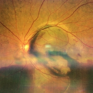
ARMD, SRNVM with sub retinal hemorrhage, 79-year-old white female, 20/300.
Condition/keywords: neovascular age-related macular degeneration (AMD), subretinal hemorrhage
-
 ARMD, SRNVM
Aug 7 2013 by H. Michael Lambert, MD
ARMD, SRNVM, with sub retinal hemorrhage: 79-year-old white female, 20/300 .
Condition/keywords: subretinal hemorrhage, subretinal neovascularization (SRNV)
-
 Choroidal Rupture with Subretinal Hemorrhage
Oct 18 2017 by Theodore Leng, MD, MS, FASRS
Choroidal rupture with subretinal hemorrhage after tennis ball injury to the eye.
Condition/keywords: choroidal rupture, subretinal hemorrhage
-
 Choroidal Tear-Multimodal imaging
Sep 10 2023 by Maneesh M Bapaye, MD, MBA
Multimodal imaging for a 26 years old patient with history of blunt trauma. Color fundus photo shows choroidal tear through foveal center with subretinal haem, autofluorescence image shows hypoAF demarkating margins and extent of the tear while the OCT B-Scan through foveal center shows tear in choroid, BM and RPE along with elevated EZ parallel to fovea with underlying hyper reflectivity s/o hemorrhage
Photographer: Maneesh Bapaye
Condition/keywords: blunt trauma, choroidal tear, subretinal hemorrhage
-
 Coats' Disease
Nov 30 2018 by Darin R. Goldman, MD
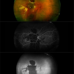
58-year-old male with a Coats’-like process in his right eye. The patient has undergone both laser photocoagulation and anti-VEGF therapy.
Photographer: Harold Rodriguez, CMA, Retina Group of Florida
Imaging device: Optomap color image, fluorescein angiogram, and fundus autofluorescence
Condition/keywords: Coats' disease, macroaneurysm, serous retinal detachment, subretinal hemorrhage
-
 Color Montage Picture
Jan 15 2019 by Aniruddha Maiti, MBBS DO DNB FRVS FICO MRCS FACS FASRS FRCOphth
Dilated tortuous vessels with intraretinal and subretinal hemorrhage at fovea.
Photographer: Sangeeta Mohanta
Imaging device: Zeiss FF450 plus IR
Condition/keywords: dilated tortuous vessels, subretinal hemorrhage, Wyburn-Mason
-
 Combined Hamartoma
Feb 29 2016 by Andrea Arriola-Lopez, MD MSc
40 year-old man with diminished VA since 6 month ago. Fundus examination revealed macular folds, yellow-whitish elevated lesion at the fovea and a subretinal hemorrhage.
Photographer: Andrea Elizabeth Arriola-Lopez MD, MSc
Imaging device: OPTOS Dakota
Condition/keywords: combined hamartoma, macula, subretinal hemorrhage
-
 Exudative Age-Related Macular Degeneration
Nov 5 2019 by Nichole Lewis
84-year-old female with exudative macular degeneration, subretinal hemorrhage and subretinal fluid.
Photographer: NIchole Lewis
Imaging device: Optos
Condition/keywords: exudative age-related macular degeneration, subretinal fluid, subretinal hemorrhage, wet age-related macular degeneration (wet AMD)
-
 FA 40 Seconds - Large Hemorrhage With Macular Detachment Due to AMD
Nov 7 2019 by John S. King, MD
81-year-old white female with three day history of seeing a "dark blob" nasally OD; no blood thinners; vision was 20/100- J16 with 2+NSC OD; OCT (not shown) had large SRF that included the fovea and extended out temporally. Posterior segment showed a large amount of SRF in the macula with some SRH in the inferior portion of the macula, hemorrhagic PEDs temporally with some RPE scarring and SRH in the periphery. On the FA there is blockage by the SRH and SRPE heme; there is staining peripherally; there is a wave of leakage that extends out into the macula and pools into to subretinal space.
Photographer: Brandon Peter
Condition/keywords: retinal pigment epithelium, subretinal hemorrhage, wet age-related macular degeneration (wet AMD)
-
 FA 5 min - Large Hemorrhage With Macular Detachment Due to AMD
Nov 7 2019 by John S. King, MD
81-year-old white female with three day history of seeing a "dark blob" nasally OD; no blood thinners; vision was 20/100- J16 with 2+NSC OD; OCT (not shown) had large SRF that included the fovea and extended out temporally. Posterior segment showed a large amount of SRF in the macula with some SRH in the inferior portion of the macula, hemorrhagic PEDs temporally with some RPE scarring and SRH in the periphery. On the FA there is blockage by the SRH and SRPE heme; there is staining peripherally; there is a wavbe of leakage that extends out into the macula and pools into to subretinal space.
Photographer: Brandon Peter
Condition/keywords: retinal pigment epithelium, subretinal hemorrhage, wet age-related macular degeneration (wet AMD)
-
 Hemorrhagic Pigment Epithelial Detachment
Dec 14 2016 by Hashim Ali Khan, OD, FAAO
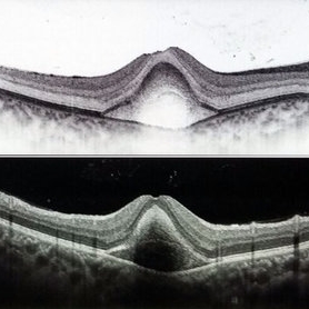
OCT of a 20-year-old female after trauma with tennis-ball, showing a hemorrhagic PED. RPE is elevated. The second Hyper-reflective band corresponding to Bruchs membrane (BM complex) is visible.
Condition/keywords: pigment epithelial detachment (PED), subretinal hemorrhage
-
 Large Subretinal Bleed in Case of Wet ARMD
Sep 28 2024 by Anjana Mirajkar, MS Ophthalmology
An intra operative image showing large sub retinal hemorrhage involving the macular area and along the superior arcade with exudation at the macular area in case of wet ARMD.
Photographer: Dr. Anjana Mirajkar -Retina Foundation, Ahmedabad
Condition/keywords: polypoidal choroidal vasculopathy (PCV), subretinal hemorrhage, wet age-related macular degeneration (wet AMD)
-
 Massive SRH in Patient on Coumadin Being Treated for Exudative AMD
Sep 30 2019 by John S. King, MD
78-year-old white female using 1mg of warfarin for atrial fibrillation, who had a large PED, Type 1 lesion from AMD. Noticed acute darkening of vision one week after anti-VEGF injection. Has very large SRH, subRPE heme, and corrugated retinal appearance post RPE tear. Vision HM (from 20/100). 20/25 in the fellow eye that has dry AMD.
Photographer: Shelly Blair
Imaging device: Optos CA
Condition/keywords: subretinal hemorrhage, wet age-related macular degeneration (wet AMD)
-
 Massive Subretinal Hemorrhage
Aug 12 2022 by Sashwanthi Mohan
A 70 year old woman with a large area of subretinal hemorrhage secondary topolychoroidal vasculopathy
Photographer: Leo John
Condition/keywords: CNVM, subretinal hemorrhage
-
 Neovascular Age-Related Macular Degeneration (1)
Apr 28 2021 by Ambar Faridi, MD
80-year-old woman with neovascular age-related macular degeneration with large subretinal hemorrhage, hemorrhagic PED, and vascular lipid exudation.
Photographer: Jennifer Tu-Bui, VA Portland Health Care System
Condition/keywords: subretinal fibrosis, subretinal hemorrhage
-
 Optos Picture 1 month after PPV with sub retinal tpa
Apr 27 2023 by Jesus Lozano, MD
Optos Image POM1 after Vitrectomy + Subretinal tPA do to subretinal hemorrhage at the macula. Visual Improvement to 6/24.
Imaging device: Optos
Condition/keywords: subretinal hemorrhage, vitrectomy
-
 PCV Polypoidal Choroidal Vasculopathy
Feb 19 2022 by Vishal Gupta, MBBS, MS
Massive Subretinal Haemorrhage and PEDs in a 62 year old female patient.
Photographer: Dr Shobhit Chawla, Prakash Netra Kendr, Lucknow, UP, INDIA
Imaging device: Zeiss Clarus 500
Condition/keywords: hemorrhagic PED, polypoidal choroidal vasculopathy (PCV), subretinal hemorrhage
-
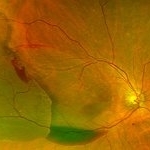 Photo of Large Hemorrhage with macular detachment due to AMD
Nov 7 2019 by John S. King, MD
81-year-old white female with three day history of seeing a "dark blob" nasally OD; no blood thinners; vision was 20/100- J16 with 2+NSC OD; OCT (not shown) had large SRF that included the fovea and extended out temporally. Posterior segment showed a large amount of SRF in the macula with some SRH in the inferior portion of the macula, hemorrhagic PEDs temporally with some RPE scarring and SRH in the periphery. On the FA there is blockage by the SRH and SRPE heme; there is staining peripherally; there is a wavbe of leakage that extends out into the macula and pools into to subretinal space. Anti-VEGF given; f/u one month.
Photographer: Brandon Peter
Condition/keywords: retinal pigment epithelium, subretinal hemorrhage, wet age-related macular degeneration (wet AMD)
-
 Ruptured Macroaneurysm
May 22 2019 by Nichole Lewis
FA of a 91-year-old woman with a ruptured macroaneurysm, intraretinal hemorrhage and subretinal hemorrhage. VA 20/400.
Photographer: Nichole Lewis
Condition/keywords: intraretinal hemorrhage, ruptured macroaneurysm, subretinal hemorrhage
-
 Subretinal BSS and air
Apr 12 2022 by Nassim Alejandro Abreu Arbaje, MD
67 year old female who presented with complaints of 5 days of decreased vision of her left eye. She underwent PPV + BSS and Air injection in the subretinal space
Photographer: Nassim Abreu, Dr. Elias Santana Hospital
Imaging device: Ngenuity 3D system screenshot
Condition/keywords: subretinal hemorrhage
-
 Subretinal Hemorrhage
Jan 7 2020 by Stacie Neview
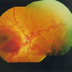
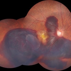
Optos fundus photograph of a 74-year-old male with severe subretinal hemorrhage and exudative retinal detachment secondary to peripheral choroidal neovascular membrane.
Photographer: Stacie Neview, Retina Specialists of Michigan, Grand Rapids Michigan, USA
Imaging device: Optos California
Condition/keywords: exudative retinal detachment, subretinal hemorrhage
-
 Subretinal Hemorrhage
Dec 2 2019 by Kristen Wagner
Fundus photo of a very large subretinal hemorrhage.
Photographer: Kristen Wagner, COT, OSC, Ophthalmic Photographer, Tennessee Retina
Condition/keywords: hemorrhage
-
 Subretinal Hemorrhage
Feb 4 2018 by Purva Patwari
65-year-old female with sudden loss of vision.
Photographer: Dr Purva Patwari
Condition/keywords: subretinal hemorrhage
 Loading…