Search results (206 results)
-
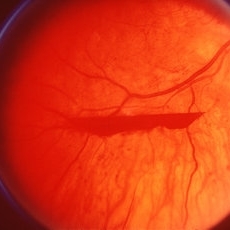
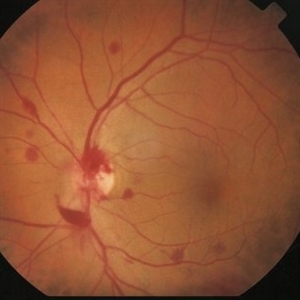
 "Boat-Shaped" Preretinal Hemorrhage
Feb 21 2019 by Mitzy E Torres Soriano, MD
Color fundus photograph showing preretinal (subhyaloid) hemorrhage in a diabetic patient with proliferative diabetic retinopathy.
Photographer: Andrea Vitale, MD
Condition/keywords: preretinal hemorrhage, proliferative diabetic retinopathy (PDR), subhyaloid hemorrhage
-
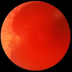 Acute Subhyaloid Hemorrhage CF
Oct 1 2012 by Jeffrey G. Gross, MD, FASRS
Acute subhyaloid hemorrhage CF.
Condition/keywords: acute, subhyaloid hemorrhage
-
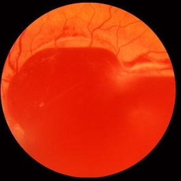 Acute Subhyaloid Hemorrhage CF
Oct 1 2012 by Jeffrey G. Gross, MD, FASRS
Acute subhyaloid hemorrhage CF.
Condition/keywords: demarcation line, subhyaloid hemorrhage
-
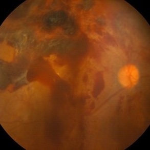
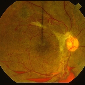 Advanced PDR
Mar 29 2013 by Henry J. Kaplan, MD
Large FPD, FPE, NVE and multiple boat shaped subhyaloid hemorrhages inferiorly, ischemic retina and multiple occluded sclerotic vessels.
Condition/keywords: foveal photoreceptor defect, subhyaloid hemorrhage
-
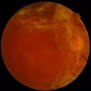 Advanced PDR
Sep 1 2014 by Hamid Ahmadieh, MD
Color fundus photograph of the right eye of a 50-year-old woman with advanced PDR.
Photographer: Soodabeh Fooladian, Negah Eye Center, Tehran, Iran
Condition/keywords: color fundus photograph, proliferative diabetic retinopathy (PDR), subhyaloid hemorrhage
-
 Anaemic Retinopathy
May 8 2023 by Akansha Sharma
Colour fundus photograph of a 38 year old male with anaemic retinoathy
Photographer: Dr. Urmil Shah, Dr. Denish Patel, Dr. Akansha Sharma, Bharati Eye Clinic, Ahmedabad, Gujarat
Condition/keywords: anaemic retinopathy, flame shaped retinal hemorrhage, subhyaloid hemorrhage
-
 Before & After - YAG Laser Hyaloidotomy for Subhyaloid Hemorrhage
Sep 19 2021 by Jesus Lozano, MD
23 year-old man with thrombocytopenia after chemotherapy d/t blastic plasmacytoid dendritic cell neoplasm. Developed a subhyaloid hemorrhage, and was treated with YAG Laser Hyaloidotomy.
Photographer: Yair Bet Yosef, Hadassah Medical Center. Israel
Imaging device: Optos
Condition/keywords: subhyaloid hemorrhage, vitreous hemorrhage
-
 Before & After - YAG Laser Hyaloidotomy for Subhyaloid Hemorrhage
Sep 19 2021 by Jesus Lozano, MD
23 year-old man with thrombocytopenia after chemotherapy d/t blastic plasmacytoid dendritic cell neoplasm. Developed a subhyaloid hemorrhage, and was treated with YAG Laser Hyaloidotomy.
Photographer: Yair Bet Yosef, Hadassah Medical Center. Israel
Imaging device: Optos
Condition/keywords: Dendritic cell Neoplasm, hyaloidotomy, subhyaloid hemorrhage, thrombocytopenia, vitreous hemorrhage
-
 Branch Retinal Vein Occlusion with Acute on Chronic Subhyaloid Hemorrhage
Oct 24 2019 by Nichole Lewis
60-year-old male with a branch retinal vein occlusion and subhyaloid hemorrhage and retinal neovascularization. VA HM.
Photographer: Nichole Lewis
Condition/keywords: branch retinal vein occlusion (BRVO), retinal neovascularization, subhyaloid hemorrhage
-
 Branch Retinal Vein Occlusion with Acute on Chronic Subhyaloid Hemorrhage
Oct 24 2019 by Nichole Lewis
60-year-old male with a branch retinal vein occlusion and subhyaloid hemorrhage and retinal neovascularization. VA HM.
Photographer: Nichole
Condition/keywords: branch retinal vein occlusion (BRVO), capillary nonperfusion, retinal neovascularization, subhyaloid hemorrhage
-
 Chronic Sub-Hyaloid Hemorrhage with Dehemoglobinized Blood
Jul 11 2025 by Aditya S Kelkar, MS, FRCS, FASRS,FRCOphth
Fundus photograph of an 38-year-old man with a long standing sub hyaloid hemorrhage with dehemoglobinized blood.
Photographer: Optom Salomi Sonawane, National Institute of Ophthalmology, Pune, India
Imaging device: Optos Daytona
Condition/keywords: chronic, dehemoglobinized hemorrhage, SUBHYALOID HEMORRHAGE
-
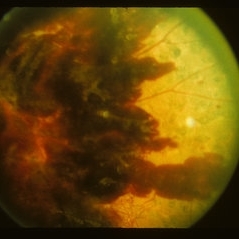
 CLOUDS OF BLOOD-Sub Hyloid Haemorrhage secondary to choroidal mass
Oct 28 2022 by Magna Mary Kuruvila
Fundus photograph of a 50 year old male patient with sudden loss of vision showing sub hyloid hemorrhage secondary to choroidal mass visualised by bscan.
Photographer: Magna Mary Kuruvila
Condition/keywords: B scan ultrasound, choroidal tumor, subhyaloid hemorrhage
-
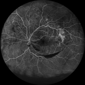 Fluorescein Angiography Neovascularization Elsewhere and Subhyaloid Hemorrhage
Aug 15 2021 by ASRS Staff
38 year-old male, presented with complaint of dark spot in vision of left eye. His vision was 6/6 in both eyes. On examination he was having subhyaloid hemorrhage and NVE in left eye.NVE was also present in RE. Patient was referred for carotid Doppler and cardiologist opinion.
Imaging device: Nidek Mirante
Condition/keywords: neovascularization elsewhere (NVE), subhyaloid hemorrhage
-
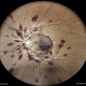
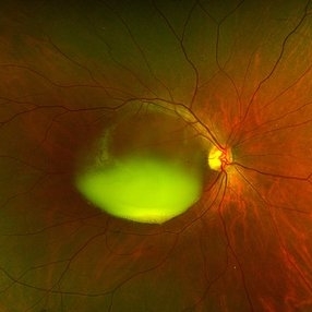
 High Risk Proliferative Diabetic Retinopathy with Sub-hyaloid Hemorrhage
May 13 2025 by Anupama Kiran Kumar
This image shows a case of high risk proliferative diabetic retinopathy. The retina is unlasered with a taut posterior hyaloid and a sub-hyaloid hemorrhage at the macula and along the arcades ,sparing the fovea.
Photographer: Mr Pratap
Imaging device: Mirante SLO/OCT (Nidek Co., Gamagori, Japan)
Condition/keywords: Diabetes, Diabetic Retinopathy, proliferative diabetic retinopathy (PDR), subhyaloid hemorrhage
-
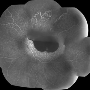
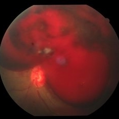
 High-Risk Proliferative Diabetic Retinopathy
Mar 20 2019 by Anfisa Ayalon, MD
Fundus fluorescein angiography of 58-year-old patient with left eye high-risk proliferative diabetic retinopathy. Note severe ischemia of retina, large areas of neovascularization elsewhere and preretinal hemorrhages.
Photographer: Anfisa Ayalon,MD., Meir Medical Center, Kfar Saba, Israel.
Imaging device: California, Optos 200 DTX
Condition/keywords: ischemia, neovascularization elsewhere (NVE), proliferative diabetic retinopathy (PDR), retina, subhyaloid hemorrhage
-
 HRC-PDR
May 2 2013 by Henry J. Kaplan, MD
Boat shaped subyaloid hemorrhage due to underlying NVE in a patient with HRC-PDR.
Condition/keywords: HRC-PDR, subhyaloid hemorrhage
-
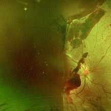
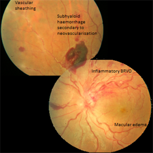 Inflammatory BRVO, Neovascularization and Macular Edema in a Patient with Vasculitis
Apr 4 2016 by AASHRAYA KARPE, MBBS, MS, FMRF
Composite fundus photograph of the left eye of a 20-year-old male with history of blurring of vision. Left eye revealed disc edema with a dilated tortuous superotemporal veins and hemorrhages in that quadrant. Mulitple preretinal hemorrhages were seen nasally along with vascular sheathing.
Imaging device: Zeiss FF450plus Fundus Camera
Condition/keywords: branch retinal vein occlusion (BRVO), macular edema, neovascularization (NV), subhyaloid hemorrhage, vascular sheathing of retina
-
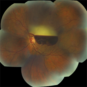
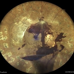
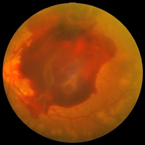
 Large Subhyaloid Hemorrhage from PDR
Jan 1 2013 by John T. Thompson, MD
Large subhyaloid hemorrhage from PDR, eye developed severe traction RD when lost to followup after this photo.
Condition/keywords: subhyaloid hemorrhage
-
 Lasered Profilerative Diabetic Retinopathy
May 8 2023 by Akansha Sharma
COLOUR FUNDUS PHOTOGRAPH OF A 42 YEAR OLD MALE WITH SUBHYALOID HEMORRHAGE IN A CASE OF LASERED PROLIFERATIVE DIABETIC RETINOPATHY
Photographer: Dr. Urmil Shah, Dr. Denish Patel, Dr. Akansha Sharma, Bharati Eye Clinic, Ahmedabad, Gujarat
Condition/keywords: PDR, proliferative diabetic retinopathy (PDR), subhyaloid hemorrhage
-
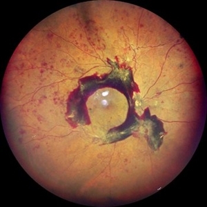
 Macroaneurysm
Jul 11 2016 by Manish Nagpal, MD, FRCS (UK), FASRS
Fundus photography of a 45 year old male who presented with a macroaneurysm in his right eye with a spill over de haemoglobinised sub hyaloid haemorrhage
Photographer: Pooja Barot
Condition/keywords: macroaneurysm, subhyaloid hemorrhage
-
 Monocular Proliferative Diabetic Retinopathy
Sep 8 2021 by VERONICA ADRIANA ROMERO- MORALES, MD
Fundus photograph of a 37-year-old woman with proliferative diabetic retinopathy and subhyaloid hemorrhage, 1 week of evolution.
Photographer: Belgica Copado Andrade
Imaging device: Cobra HD
Condition/keywords: neovascularization (NV), proliferative diabetic retinopathy (PDR), subhyaloid hemorrhage, thickening of the posterior hyaloid, vitreous blood
-
 PDR
PDR
Feb 25 2025 by Parnian Arjmand, MD, MSc, FRCSC, DABO
Young patient with proliferative diabetic retinopathy and subhyaloid hemorrhage. Extensive neovascular fronds can be noted throughout the posterior pole and disc.
Condition/keywords: subhyaloid hemorrhage
-
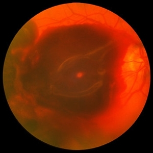 PDR with Severe Subhyaloid Hemorrhage
Oct 8 2012 by Jeffrey G. Gross, MD, FASRS
PDR with severe subhyaloid hemorrhage sparing fovea CF.
Condition/keywords: subhyaloid hemorrhage
-
 PDR with Severe Subhyaloid Hemorrhage
Oct 8 2012 by Jeffrey G. Gross, MD, FASRS
PDR with severe subhyaloid hemorrhage.
Condition/keywords: subhyaloid hemorrhage
-
 Posterior Vitreous Detachment
Aug 23 2012 by Gabriela Lopezcarasa Hernandez, MD
Subhyaloid hemorrhage secondary to posterior vitreous detachment
Photographer: Gabriela Lopezcarasa Hernandez, Hospital Angeles Lomas
Imaging device: Zeiss FF4
Condition/keywords: subhyaloid hemorrhage, vitreous detachment
 Loading…