Search results (141 results)
-
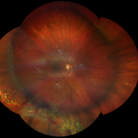
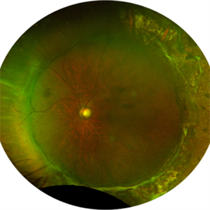
 1 year Follow Up after Scleral Buckle Surgery in a Young Patient
May 18 2023 by Jesus Lozano, MD
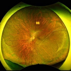
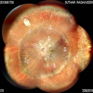
25 year old man after Scleral Buckle Surgery + laser Retinopexy do to RRD macula off with ínfero temporal mid peripheral retinal holes in an area of lattice degeneration. Final VA 6/9.
Imaging device: Optos
Condition/keywords: scleral buckle
-
 Aphakic Retinal Detachment
Nov 9 2012 by Norman Byer
This 75-year-old woman had a scleral buckling operation on this aphakic eye two months previously. Two months following surgery she again had new symptoms of blurred vision and was found to have a redetachment caused by this small tractional retinal tear which may have been missed at the time of her original operation.
Condition/keywords: aphakic eye, scleral buckle, tractional retinal tear
-
 Attached Retina in a Silicon Oil Filled Buckled Eye with Retinectomy
Apr 17 2021 by Navneet Mehrotra, DNB
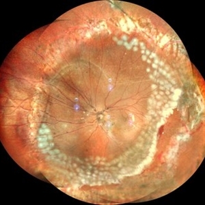
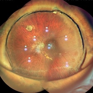
Fundus photograph of a 12-year-old boy operated for re retinal detachment with PVR showing attached retina with fresh and old laser marks, silicon oil filled and relaxing retinectomy.
Photographer: Dr Nivesh Gupta, Retina Foundation
Imaging device: Nidek mirante
Condition/keywords: proliferative vitreoretinopathy (PVR), retinectomy, scleral buckle
-
 Branch Retinal Vein Occlusion
Dec 9 2020 by Olivia Rainey
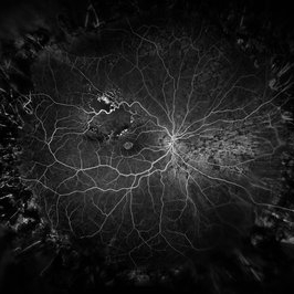
Ultra-widefield angiogram of a 78-year-old male with a branch retinal vein occlusion affecting his right eye. The patient was diagnosed on 12/17/12 at another practice. The physician noted that there wasn't NVE noted, however areas of micoaneurysmal dilation is present. She noted retinal ischemia secondary to BRVO. 12/8/20 leakage on FA noted to be worsening compared to his previous angiography. She has concern for progressing NVE and recommends sector PRP after injection of antiVEGF series of 3 for the health of the eye.
Photographer: Olivia Rainey, OCT-C, COA
Imaging device: Optos California
Condition/keywords: branch retinal vein occlusion (BRVO), macular branch retinal vein occlusion (BRVO), non-perfusion, scleral buckle, vitreoretinal surgery
-
 Buckle intrusion with Retinal detachment
Feb 8 2018 by Manish Nagpal, MD, FRCS (UK), FASRS
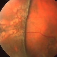
Patient operated on 10 years back for a scleral buckling surgery presented with decreased vision and had a superonasal retinal detachment along with intrusion of the scleral buckle.
Photographer: Mehul Prajapati
Condition/keywords: acute retinal detachment, retinal break, scleral buckle
-
 Buckled Eye in a case of High Myopia
Jan 8 2020 by Sham Talati, DOMS
Buckled Eye in a case of high myopia.
Photographer: Dr. Sham Talati,Retina Foundation,Ahmedabad
Imaging device: Nidek Mirante
Condition/keywords: high myopia, scleral buckle
-
 Buckled Retinal Detachment
Aug 10 2019 by Manish Nagpal, MD, FRCS (UK), FASRS
Follow up of a patient who underwent cryo and buckling using chandelier based viewing systems.
Photographer: Gayathri Mohan, Retina Foundation
Condition/keywords: scleral buckle
-
 Buckled Silicon oil Filled eye
Jul 25 2019 by Manish Nagpal, MD, FRCS (UK), FASRS
Wide field view of a buckled eye along with silicon oil reflex.
Photographer: Gayathri Mohan, Retina Foundation
Imaging device: Nidek Mirante SLO
Condition/keywords: scleral buckle, silicone oil
-
 Bullous Retinoschisis status post Scleral Buckle
Apr 2 2019 by Gary R. Cook, MD, FACS
39-year-old white male 2 weeks status post scleral buckling for bullous retinoschisis temporally OD threatening the macula
Imaging device: Topcon VT-50
Condition/keywords: bullous retinoschisis, scleral buckle
-
 Choroidal Vessels Transillumination
Mar 20 2024 by Kingston Rodolfo Ureña-Wong, MD, Opht, MSc
Photograph of choroidal vessels transillumination during a scleral buckle to repair a complete retinal detachment.
Photographer: Garagarza-Mariscal Heber, APEC.
Condition/keywords: choroidal vessels, scleral buckle
-
 Combined Tractional and Rhegmatogenous Retinal Detachment
Jan 30 2023 by Olivia Rainey
Ultra-widefield fluorescein angiography of a combined tractional and rhegmatogenous retinal detachment repair affecting the left eye. The retina is attached following silicone oil placement during most recent surgery. The patient was seeing CF at the time the image was taken.
Photographer: Olivia Rainey, OCT-C, COA
Imaging device: Optos California
Condition/keywords: diabetes, diabetic macular edema, diabetic retinopathy, fluorescein angiogram (FA), hyperfluorescence, right eye, scleral buckle, silicone oil, tractional retinal detachment, ultra-wide field imaging, ultra-widefield image
-
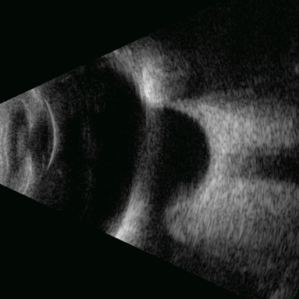 Deep Scleral Buckle
May 5 2025 by Gustavo Uriel Fonseca Aguirre
This B-mode axial ultrasound scan shows an eye with a scleral buckle in place for previous rhegmatogenous retinal detachment. The image demonstrates the characteristic indentation of the ocular wall at the buckle site, with proper retinal reattachment.
Photographer: Gustavo U. Fonseca Aguirre, Hospital Conde de Valenciana, Ciudad de México
Condition/keywords: scleral buckle
-
 Dialysis Repair with Scleral Buckle
May 7 2023 by Maxwell J Wingelaar, MD
A 28 year old male with a retinal dialysis that was repair successfully with a scleral buckle
Photographer: Ken Huff
Condition/keywords: retinal dialysis, scleral buckle
-
 Encircling Scleral Buckle Axial View
Dec 10 2012 by Yale L. Fisher, MD
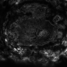
The buckle is an encircling element. the anterior posterior view shows a cross section above and below on the screen. Rotation vertically demonstrates a superior and inferior cross-section of a highly elevated scleral buckle. There is movement of the separated formed vitreous and far anterior (visible at the left of the screen). Optic nerve shadow is visible as the nerve moves vertically.
Condition/keywords: B scan ultrasound, scleral buckle, video
-
 Erosion of Segmental Buckle
Feb 25 2022 by Roger A. Goldberg, MD, MBA
Erosion of sharp edge of segmental scleral buckle seen 15 years after being placed for repair of a retinal detachment
Photographer: Melissa Bartlett, Bay Area Retina Associates
Imaging device: Optos
Condition/keywords: retinal defect, scleral buckle
-
 Erroding Buckle
Mar 1 2017 by Philip J. Polkinghorne, MD
Fundus photograph of an eroding buckle. The clinical signs have not changed in 5 years.
Photographer: Philip Polkinghorne
Condition/keywords: complication, scleral buckle
-
 Exposed Scleral Buckle, Infection - Temporal View
Feb 4 2013 by James B. Soque, CRA, OCT-C, COA, FOPS
External Photograph of a 66-year-old WM with Hx of SBOD in 2009, graft attempt failed, infection resulted. Scheduled for removal of SBOD.
Photographer: James Soque CRA COA
Imaging device: External Photo, Topcon TRC 50 DX, MERGE software
Condition/keywords: scleral buckle, suture exposed
-
 Exposed Scleral Buckle, with Exposed Suture, Infection - Infero Nasal View, Upgaze
Feb 4 2013 by James B. Soque, CRA, OCT-C, COA, FOPS
External Photograph of a 66-year-old WM with Hx of SBOD in 2009, graft attempt failed, infection resulted. Scheduled for removal of SBOD.
Photographer: James Soque CRA COA
Imaging device: External Photo, Topcon TRC 50 DX, MERGE software
Condition/keywords: scleral buckle, suture exposed
-
 Exposed Scleral Buckle, with Exposed Suture, Infection - Infero Temporal View
Feb 4 2013 by James B. Soque, CRA, OCT-C, COA, FOPS
External Photograph of a 66-year-old WM with Hx of SBOD in 2009, graft attempt failed, infection resulted. Scheduled for removal of SBOD.
Photographer: James Soque CRA COA
Imaging device: External Photo, Topcon TRC 50 DX, MERGE software
Condition/keywords: exposed suture, scleral buckle
-
 Exposed Scleral Buckle, with Infection - Infero Nasal View
Feb 4 2013 by James B. Soque, CRA, OCT-C, COA, FOPS
External photograph of a 66-year-old WM with Hx of SBOD in 2009, graft attempt failed, infection resulted. Scheduled for removal of SBOD.
Photographer: James Soque CRA COA
Imaging device: External Photo, Topcon TRC 50 DX, MERGE software
Condition/keywords: scleral buckle, suture exposed
-
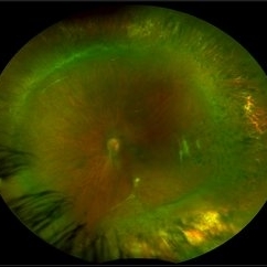 Familial Exudative Vitreoretinopathy
Feb 2 2018 by Olivia Rainey
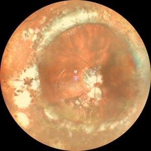
Ultra-wide field montage of a 37-year-old female with familial exudative vitreoretinopathy affecting her left eye. Cryotherapy, laser destruction of retinopathy, and a scleral buckle was performed to stabilize the retina in 2017.
Photographer: Olivia Rainey
Imaging device: Optos
Condition/keywords: familial exudative vitreoretinopathy (FEVR), fibrotic neovascularization, laser scarring, left eye, montage, Optos, scleral buckle, tractional retinal detachment, ultra-wide field imaging
-
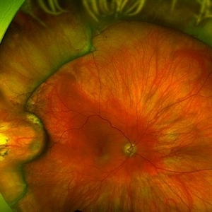
 Fully reattached retina following scleral buckling with a 360 encircling band and supero temporal buckle in a 25 year old woman.
Nov 9 2023 by Jesus Lozano, MD
Optos image of a 25 year old woman with a fully reattached retina following scleral buckling with a 360 encircling band and supero temporal buckle
Photographer: Mihaela Shlomi, Hillel Yaffe Medical Center. Israel.
Imaging device: Optos
Condition/keywords: scleral buckle
-
 Giant Retinal Tear After Successful Re-Attachment
Jun 12 2023 by Ethan K Sobol, MD
Appearance of a giant retinal tear eight months after a successful scleral buckle with vitrectomy and silicone oil tamponade, followed by silicone oil removal. Central haziness in the image is due to the development of a cataract.
Condition/keywords: GRT, scleral buckle
-
 Horseshoe Tear
Jun 24 2015 by Andree Henaine-Berra, MD
Photograph of the right eye of a 58-year-old male patient with a retinal detachment due to a peripheral horseshoe tear, showing the moment when cryotherapy is applied during the scleral bluckling procedure.
Photographer: Jorge Morales, MD. Hospital General "Dr. Manuel Gea Gonzalez". Mexico City.
Condition/keywords: acute retinal detachment, cryotherapy, scleral buckle
-
 Horseshoe Tear With Scleral Buckle
Oct 31 2013 by Jason S. Calhoun
Patient had a retinal detachment with retinal tear superior temporally. Underwent surgery and had a scleral buckle placed with good support of the tear. VA is count fingers and will return in 2-months for follow up.
Photographer: Jason S. Calhoun, Ophthalmic Photographer, Department of Ophthalmology, Mayo Clinic Jacksonville
Imaging device: TOPCON TRC 50-EX
Condition/keywords: retinal tear, scleral buckle
 Loading…