Search results (138 results)
-
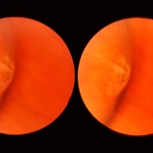
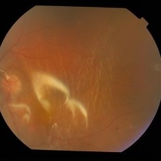
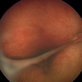 Congenital Retinal Fold
Feb 9 2017 by Dominic M Buzzacco, MD
Fundus photograph of10-month-old male with congenital retinal fold. Normal contralateral eye.
Photographer: Dominic M Buzzacco MD, Midwest Retina
Imaging device: Retcam3
Condition/keywords: retinal fold
-
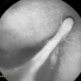
 Fish Hook Eye Trauma
Jun 12 2024 by Miguel Brito, MD, FASRS
Fundus photograph of a 15-year-old boy post cataract aspiration, pars plana vitrectomy, suprachoroidal drainage, and retinal reattachment surgery secondary to traumatic endophthalmitis.
Photographer: Miguel Brito
Condition/keywords: endophthalmitis, PFCL, Retinal detachment under Silicon Oil, retinal fold
-
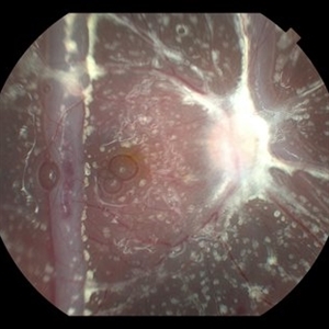
 Giant Retinal Tear With Retinal Fold
Jun 13 2024 by Anand Temkar
Intraoperative still of a 34 year old male showing giant retinal tear with retinal fold.
Photographer: Dr.Anand Temkar- Retina Foundation, Ahmedabad
Condition/keywords: giant retinal tear, GRT, retinal fold
-
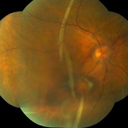
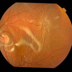
 Large Retinal Fold
Sep 22 2020 by Sophia El Hamichi, MD
A 69-year-old female, with a history of choroidal melanoma in her left eye with exudative detachment, underwent tumor laser ablation. She then developed a complex combined tractional and rhegmatogenous retinal detachment with a giant retinal tear. The patient underwent surgical repair of her retinal detachment with pars plana vitrectomy and silicone oil. In the post-op, the patient developed large retinal folds masking the optic nerve depicted in the fundus photograph.
Photographer: Belinda Rodriguez, Murray Ocular Oncology and Retina, Miami
Condition/keywords: melanoma, pars plana vitrectomy (PPV), retinal fold, silicone oil
-
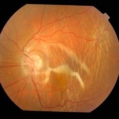
 Large Retinal Fold Masking the Optic Nerve
Sep 22 2020 by Sophia El Hamichi, MD
A 69-year-old female, with a history of choroidal melanoma in her left eye with exudative detachment, underwent tumor laser ablation. She then developed a complex combined tractional and rhegmatogenous retinal detachment with a giant retinal tear. The patient underwent surgical repair of her retinal detachment with pars plana vitrectomy and silicone oil. In the post-op, the patient developed large retinal folds masking the optic nerve depicted in the OCT photograph.
Photographer: Belinda Rodriguez, Murray Ocular Oncology and Retina, Miami
Condition/keywords: giant retinal tear, melanoma, pars plana vitrectomy (PPV), retinal fold, silicone oil
-
 Macular Fold Following Retinal Detachment Surgery
May 30 2014 by Mitzy E Torres Soriano, MD
Fundus photograph of a 38-years old man with macular fold following retinal detachment surgery.
Photographer: Ricardo Montoya. Asociación para evitar la Ceguera. México.
Condition/keywords: macular fold, post-op, retinal fold
-
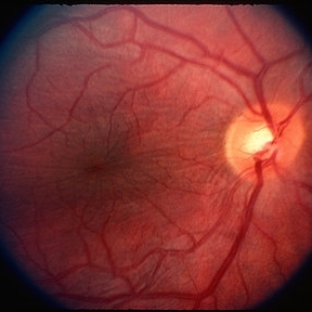
 Meridional Fold
Nov 9 2012 by Norman Byer
This is the same lesion as in the previous photograph. With the scleral indentation placed more posterior, we now can see that the fold ends over a small collection of subretinal fluid and that there is a very tiny retinal hole just below the posterior end of the retinal fold.
Condition/keywords: peripheral cystoid degeneration, retinal fold, retinal hole, scleral indentation, subretinal fluid
-
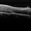
 Posterior Retinal Folds
Feb 9 2015 by Leandro C. Zacharias, MD, PhD
Fundus photograph of a 59-year-old woman 3 weeks after buckle for a macula-off retinal detachment.
Photographer: Leandro Cabral Zacharias
Imaging device: Zeiss Visucam
Condition/keywords: retinal fold
-
 Proliferative Vitreoretinopathy
Mar 3 2017 by Nichole Lewis
62-year-old female s/p retinal detachment repair with proliferative vitreoretinopathy inferior with early star folds.
Photographer: Nichole Lewis
Condition/keywords: proliferative vitreoretinopathy (PVR), retinal fold
-
 Retinal Fold
Mar 9 2015 by Matt Poe, COA
This patient developed a retinal fold following a retinal detachment repair. The patient underwent another retinal detachment surgery to fix the retinal fold. The patient's retina was fixed and did well post-operative.
Photographer: Matt Poe, COA. Northwest Arkansas Retina Associates, Springdale, AR.
Condition/keywords: optical coherence tomography (OCT), retinal fold
-
 Retinal Fold
Sep 26 2023 by Mauricio Bayram-Suverza, MD
A 38-year-old man underwent vitrectomy in the left eye due to a giant tear in the upper retina. SF6 gas was used as endotamponade. During the post-surgical check-up, it was identified that the patient developed a full-thickness retinal fold due to retinal slippage during fluid-air exchange. As the fold was away from the macular area, it was decided to observe the patient. Three weeks after the surgery, his best-corrected visual acuity was 20/30.
Photographer: Mauricio Bayram-Suverza, Fundación Hospital Nuestra Señora de la Luz
Imaging device: TRC-50DX
Condition/keywords: giant retinal tear, retina surgery complications, Retinal slippage, vitreoretinal surgery
-
 Retinal Fold Angiography
Feb 9 2017 by Dominic M Buzzacco, MD
Late phase angiogram of 10-month-old male with congenital retinal fold. Contralateral eye had normal angiography.
Photographer: Dominic M Buzzacco MD, Midwest Retina
Imaging device: Retcam 3
Condition/keywords: retinal fold
-
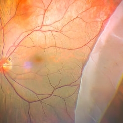
 Retinal Fold Near Retinal Break in Rhegmatogenous Retinal Detachment
Dec 15 2014 by Mallika Goyal, MD
Left fundus of a 32-year-old male shows a large retinal break with rhegmatogenous retinal detachment. This is near a fixed retinal fold (not seen in this photograph).
Photographer: Mallika Goyal, MD, Apollo Health City, Jubilee Hills, Hyderabad-500033
Condition/keywords: retinal fold
-
 Retinal Fold Near Retinal Break in Rhegmatogenous Retinal Detachment
Dec 15 2014 by Mallika Goyal, MD
Left fundus of a 32-year-old male shows a fixed retinal fold. This is adjacent to a large retinal break (not seen here) with rhegmatogenous retinal detachment.
Photographer: Mallika Goyal, MD, Apollo Health City, Jubilee Hills, Hyderabad-500033
Condition/keywords: retinal fold
-
 Retinal Fold Near Retinal Break in Rhegmatogenous Retinal Detachment
Dec 15 2014 by Mallika Goyal, MD
Left fundus of a 32-year-old male shows a fixed retinal fold near a large retinal break with rhegmatogenous retinal detachment.
Photographer: Mallika Goyal, MD, Apollo Health City, Jubilee Hills, Hyderabad-500033
Condition/keywords: retinal fold
-
 Retinal Folds
Mar 29 2013 by Henry J. Kaplan, MD
Retinal folds as fine wrinklings.
Condition/keywords: retinal fold
-
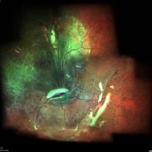
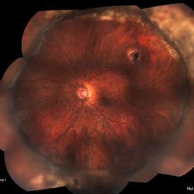
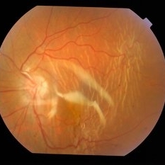
 Retinal folds following retinal reattachment surgery
Nov 22 2015 by Mallika Goyal, MD
Multiple retinal folds 4 weeks following vitreous surgery (perfluorodecalin assisted) for retinal detachment with giant retinal tear.
Photographer: Mallika Goyal, MD, Apollo Health City, Jubilee Hills, Hyderabad, India
Condition/keywords: retinal fold
-
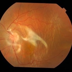
 Retinal Folds Following Retinal Reattachment Surgery
Nov 22 2015 by Mallika Goyal, MD
Multiple retinal folds 4 weeks following vitreous surgery (perfluorodecalin assisted) for retinal detachment with giant retinal tear.
Photographer: Mallika Goyal, MD, Apollo Health City, Jubilee Hills, Hyderabad, India
Condition/keywords: retinal fold
-
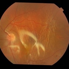
 Retinal Folds Following Retinal Reattachment Surgery
Nov 22 2015 by Mallika Goyal, MD
Multiple retinal folds 4 weeks following vitreous surgery (perfluorodecalin assisted) for retinal detachment with giant retinal tear.
Photographer: Mallika Goyal, MD, Apollo Health City, Jubilee Hills, Hyderabad, India
Condition/keywords: retinal fold
-
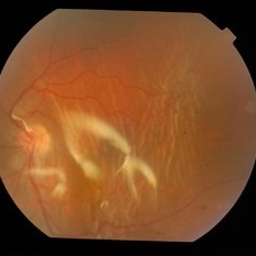
 Retinal Folds Following Retinal Reattachment Surgery
Nov 22 2015 by Mallika Goyal, MD
Multiple retinal folds 4 weeks following vitreous surgery (perfluorodecalin assisted) for retinal detachment with giant retinal tear.
Photographer: Mallika Goyal, MD, Apollo Health City, Jubilee Hills, Hyderabad, India
Condition/keywords: retinal fold
-
 Retinal Folds Following Retinal Reattachment Surgery
Nov 22 2015 by Mallika Goyal, MD
Multiple retinal folds 4 weeks following vitreous surgery (perfluorodecalin assisted) for retinal detachment with giant retinal tear.
Photographer: Mallika Goyal, MD, Apollo Health City, Jubilee Hills, Hyderabad, India
Condition/keywords: retinal fold
 Loading…