Search results (66 results)
-
 Acute Retinal Detachment
Nov 9 2012 by Norman Byer
This 54-year-old man was referred because of sudden symptoms in his opposite eye in which he had suffered an acute retinal detachment secondary to a horseshoe tear around lattice degeneration. During the examination, the fellow eye shown here was also found to have this large horseshoe tear about 1 o’clock hour (4 disc diameters) in size. A tear occurred around a lattice lesion which is present on the flap but is out of focus. This tear had been asymptomatic even though it was caused by a posterior vitreous detachment and illustrates that even very large tears may produce no symptoms or mild symptoms that are easily overlooked.
Condition/keywords: lattice degeneration, posterior vitreous detachment
-
 Cystic Retinal Tuft
Nov 9 2012 by Norman Byer
This is the same lesion as in the previous slide pair but the photograph was taken nine years later when the patient was 58-years-old soon after an acute posterior vitreous detachment. This demonstrates that posterior vitreous detachment can produce large retinal tears at these sites. However, it is important to emphasize that prophylactic treatment of cystic retinal tufts in the absence of a retinal tear would be very ill-advised because several hundred innocence and harmless lesions would have to be treated in order to prevent one tear of the retina.
Condition/keywords: cystic retinal tuft, posterior vitreous detachment, retinal tear
-
 ERM that Spontaneously Peeled
Oct 8 2012 by David R. Chow, MD, FRCS(C)
An ERM that through follow-up sponateously separated with the development of PVD.
Condition/keywords: epiretinal membrane (ERM), posterior vitreous detachment
-
 Evolving Weiss Ring
Sep 11 2022 by Michael B Green, MD, MBA
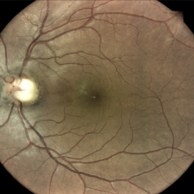
Fundus photograph of a 62-year-old female with an evolving Weiss-ring in the process of separating from the optic disc.
Condition/keywords: posterior vitreous detachment, PVD, Weiss ring
-
 Extreme Asteroid Hyalosis
Apr 27 2016 by Matt Poe, COA
This patient was sent for a possible retinal detachment. Extreme difficult view of posterior pole due to asteroid hyalosis. After B-Scan was performed it was determined patient did not have a retinal detachment, only posterior vitreous detachment.
Photographer: Matt Poe, COA. Northwest Arkansas Retina Associates, Springdale, AR.
Condition/keywords: asteroid hyalosis, posterior vitreous detachment
-
 Lattice Degeneration
Nov 9 2012 by Norman Byer
Lattice degeneration in a 42-year-old man which has produced four atrophic holes in a linear arrangement surrounded by a subclinical retinal detachment of unknown duration. By age 63, 21 years later, a posterior vitreous detachment was diagnosed in this eye, which was not present four years earlier. Nevertheless, the appearance seen here has remained exactly the same for 30 years, more than eight years with a concurrent PVD.
Condition/keywords: atrophic retinal hole, lattice degeneration, posterior vitreous detachment
-
 Morning-Glory-Syndrome
Dec 22 2017 by James B. Soque, CRA, OCT-C, COA, FOPS
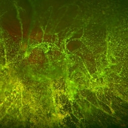
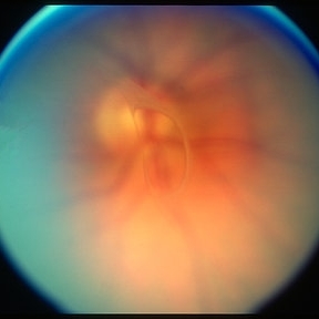
68-year-old WM with Morning Glory Syndrome with PVD OS with Staphyloma surrounding optic nerve and extending into the macula. Also, esotropia OS from V1 nerve paresis from birth, with amblyopia.
Photographer: James B Soque, CRA OCT-C COA FOPS
Imaging device: Optos Daytona
Condition/keywords: color photo, esotropia, fundus photograph, Optomap, Optos, peripheral vascular disease (PVD), posterior vitreous detachment, staphyloma, ultra-wide field imaging, wide angle imaging
-
 OCT of a Posterior Vitreous Detachment
Nov 26 2019 by Geoffrey G. Emerson, MD, PhD, FASRS
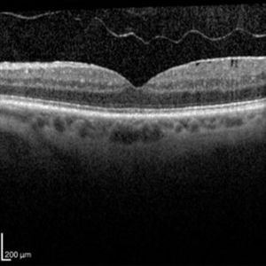
OCT of a posterior vitreous detachment.
Condition/keywords: optical coherence tomography (OCT), posterior vitreous detachment
-
 Peripapillary Glial Proliferation
Oct 18 2012 by Suber S. Huang, MD, MBA, FASRS
61-year-old woman with peripapillary gilal proliferation
Photographer: Stacie Hrvatin
Condition/keywords: glial proliferation, posterior vitreous detachment, Weiss ring
-
 Peripapillary Glial Proliferation
Oct 18 2012 by Suber S. Huang, MD, MBA, FASRS
61-year-old woman with peripapillary gilal proliferation
Photographer: Stacie Hrvatin
Condition/keywords: glial proliferation, posterior vitreous detachment, Weiss ring
-
 Peripapillary Glial Proliferation
Oct 18 2012 by Suber S. Huang, MD, MBA, FASRS
61-year-old woman with peripapillary gilal proliferation
Photographer: Stacie Hrvatin
Condition/keywords: glial proliferation, posterior vitreous detachment, Weiss ring
-
 Peripapillary Glial Proliferation
Oct 18 2012 by Suber S. Huang, MD, MBA, FASRS
61-year-old woman with peripapillary gilal proliferation
Photographer: Stacie Hrvatin
Condition/keywords: glial proliferation, posterior vitreous detachment, Weiss ring
-
 Posterior Vitreous Detachment
Aug 23 2012 by Gabriela Lopezcarasa Hernandez, MD
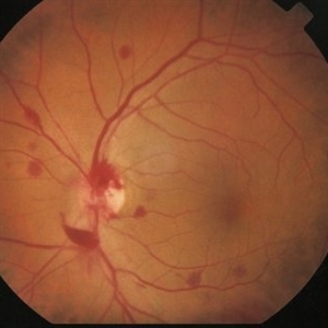
Subhyaloid hemorrhage secondary to posterior vitreous detachment
Photographer: Gabriela Lopezcarasa Hernandez, Hospital Angeles Lomas
Imaging device: Zeiss FF4
Condition/keywords: subhyaloid hemorrhage, vitreous detachment
-
 Posterior vitreous detachment
Jan 11 2013 by Alex P. Hunyor, MD
Posterior vitreous detachment with prominent Weiss ring.
Condition/keywords: posterior vitreous detachment
-
 Posterior Vitreous Detachment
Nov 9 2012 by Norman Byer
In this 50-year-old man, these two adjacent tears are separated by a narrow band of tissue but have a common flap. They were caused by a posterior vitreous detachment and they are surrounded by a localized area of detachment. This case is similar to slide pair 53.
Condition/keywords: acute posterior vitreous detachment, adjacent tears, bridge of tissue between tears, posterior vitreous detachment, retinal tear
-
 Posterior Vitreous Detachment
Nov 9 2012 by Norman Byer
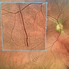
This 68-year-old woman had a recent posterior vitreous detachment which produced this symptomatic horseshoe tear exactly at the site of this cystic retinal tuft. Note the characteristic discrete white nubbin at the apex, which is produced by a cap of glial cells with densely packed cytoplasm.
Condition/keywords: cystic retinal tuft, glial cells, posterior vitreous detachment, white retinal tuft
-
 Posterior Vitreous Detachment
Mar 21 2019 by Michael Politis, MD
Intra-op image of a PVD induction using Kenalog in a retinal detachment after retained nuclear material
Photographer: Michael Politis MD, McGill University, Montreal, Canada
Imaging device: Karl Stroze
Condition/keywords: kenalog, PVD induction
-
 Posterior Vitreous Detachment
Sep 1 2020 by J. Sebag, MD, FACS, FRCOphth, FARVO
Left: Preset lens biomicroscopy of PVD in the left eye of a subject with a widely dilated pupil. The detached posterior vitreous cortex is seen (arrows) as is the optic disc and retinal vasculature (upper left). (courtesy of C. L. Trempe MD, Harvard Medical School, Boston, MA) [Sebag J: Vitreous – in Health & Disease Springer, New York, 2014; image © Springer Nature, reprinted with permission] Right: B-scan ultrasonography of PVD images the detached posterior vitreous cortex with a visible Weiss Ring.
Condition/keywords: posterior vitreous detachment
-
---thumb.jpg/image-square;max$300,300.ImageHandler) Posterior Vitreous Detachment
Posterior Vitreous Detachment
Aug 14 2013 by From the Collections of Thomas M. Aaberg, MD and Thomas M. Aaberg Jr., MD
External.
Condition/keywords: posterior vitreous detachment
-
 Posterior Vitreous Detachment
Jan 31 2025 by Thirumalesh Mochi Basavaraj, MD
Intraoperative view of Triamcinolone-assisted posterior vitreous detachment.
Photographer: Thirumalesh Mochi Basavaraj
Condition/keywords: PVD induction, triamcinolone
-
 Posterior Vitreous Detachment
Sep 28 2025 by Sanauddin Samejo , Diploma (Ophthalmic Technician Training Course)
Posterior Vitreous Detachment (PVD)
Photographer: Sanauddin Samejo
Imaging device: Optos Silver Stone
Condition/keywords: posterior vitreous detachment, PVD
-
 Posterior Vitreous Detachment
Sep 28 2025 by Sanauddin Samejo , Diploma (Ophthalmic Technician Training Course)
Posterior Vitreous Detachment (PVD)
Photographer: Sanauddin Samejo
Imaging device: Optos Silver Stone
Condition/keywords: posterior vitreous detachment, PVD
-
 Posterior Vitreous Detachment
Nov 1 2023 by ANKIT JAIN
USG B SCAN image showing membranous echoes with low to moderate spikes with free after movements with no attachment to disc suggestive of posterior vitreous detachment.
Photographer: DR ANKIT JAIN
Condition/keywords: B scan ultrasound, posterior vitreous detachment, PVD, ultrasound
-
 Retinal Tear in Aphakic Fellow Eye
Nov 9 2012 by Norman Byer
This 59-year-old man presented with sudden symptoms of retinal detachment in his opposite aphakic eye secondary to a tiny retinal tear about 1/8th disc diameter in size. During the examination, the fellow eye shown here was found to have this much larger tractional tear approximately 2 disc diameters in total length. If you look carefully, you will see that this is really a series of three separate tears with a common flap. The tears are separated by tiny bridges of remaining tissue which cause the edges of the apparent large tear to be serrated. This was also an aphakic eye with a posterior vitreous detachment but the lesion had produced no symptoms.
Condition/keywords: asymptomatic, bridge of tissue between tears, posterior vitreous detachment, tractional retinal tear
-
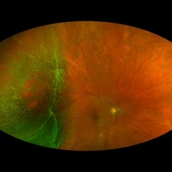 Rhegmatogenous Macula-On Retinal Detachment (Honeycomb)
Aug 6 2024 by Xitlali Caterina
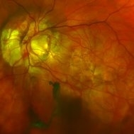
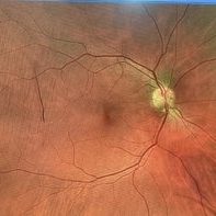
Ultra-wide field fundus photograph of a 72 year old female with a macula-on retinal detachment with multiple breaks affecting her right eye. Patient presented in the office with flashes of light for five consecutive days prior. The patients vision was sc20/30 PHNI. The physician also noted an acute posterior vitreous detachment and lattice degeneration in the affect eye.
Photographer: Xitlali Caterina
Imaging device: Optos California RGB
Condition/keywords: honeycomb, lattice degeneration, Optos, posterior vitreous detachment, Retinal Detachment with Multiple Breaks, rhegmatogenous retinal detachment, ultra-wide field imaging
 Loading…