Search results (56 results)
-
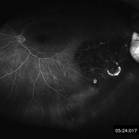
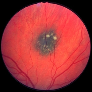
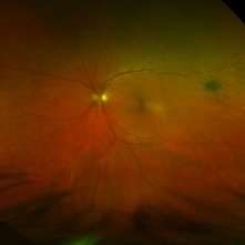
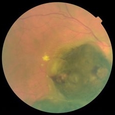 Choroidal Melanoma
May 15 2014 by Mitzy E Torres Soriano, MD
Fundus photograph of a 55-year-old male with pigmented, elevated lesion involving optic nerve. with exudation, hemorrhage and subretinal fluid.
Photographer: Mitzy E Torres Soriano, Centro Medico Cagua, Venezuela
Imaging device: Retinal camera TRC-NW8, TOPCON
Condition/keywords: choroidal nevus, pigmented lesion
-
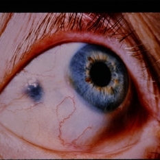
 Iris Pigmented Lesion
Apr 27 2018 by Mark Lazcano
Gonio photograph of 20-year-old male with pigmented iris lesion consistent with melanocytoma
Photographer: mark Lazcano,University of Miami , Bascom Palmer Eye Institute
Imaging device: gonio Prism
Condition/keywords: pigmented lesion
-
 Lattice Lesion
Nov 9 2012 by Norman Byer
This is a photograph of a lattice lesion in a 23-year-old girl taken without scleral indentation. Just to the left of the center of the slide is a slightly pigmented lesion almost oval in shape with a retinal hole in each end. Ten years earlier at the age of 13 this lesion appeared exactly like the one in the previous case as a pure red crater. Five years later two new round retinal holes were seen, one in each end, with a tiny bit of subretinal fluid within the lattice lesion only. Five years later still the appearance was as shown in this slide pair with the subretinal fluid now extending slightly beyond the lattice lesion as far as the curved row of tiny yellow exudates seen just to the right of the center of the slide. It is now actually a small subclinical retinal detachment. The next slide pair will show this better using scleral indentation.
Condition/keywords: lattice degeneration, lattice lesion, pigmented lesion, reddish crater, retinal hole, subretinal fluid, yellow exudate
-
 Scleral Indentation
Nov 9 2012 by Norman Byer
The next three photographs are of the same lesion in a 26-year-old man and demonstrate the value of scleral indentation. This view without indentation shows only a tiny pigmented and atrophic spot in the fundus.
Condition/keywords: atrophic spot, pigmented lesion, scleral indentation
-
 61-Year-Old Man With Large Peripheral CHRPE
Dec 9 2017 by Timothy S Fuller, MD
61-year-old man presented for evaluation of pigmented retinal lesion. Found to have a large, peripheral CHRPE with characteristic lacunae, sharp margins, and lack of elevation.
Condition/keywords: benign pigmented lesions, congenital hypertrophy of the retinal pigment epithelium (CHRPE), lacunae
-
 Adenocarcinoma Arising from CHRPE
Sep 17 2015 by Marc C. Peden, MD
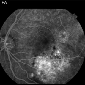
49-year-old female referred for presumed ocular melanoma. On examination was noted to have darkly pigmented lesion in the temporal retina of left eye. Lesion had characteristic scalloped edges with central lacunae, however, on ultrasonography was noted to have 1.8mm of elevation with high internal reflectivity. IVFA shows absence of dual circulation with areas of window defect. Findings were consistent with those described by Shields et al., in their April 2001 article in Archives of Ophthalmology.
Photographer: Janet Traynom
Imaging device: Optos P200MA
Condition/keywords: adenocarcinoma arising from CHRPE
-
 Adenocarcinoma Arising from CHRPE
Sep 17 2015 by Marc C. Peden, MD
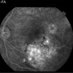
49-year-old female referred for presumed ocular melanoma. On examination was noted to have darkly pigmented lesion in the temporal retina of left eye. Lesion had characteristic scalloped edges with central lacunae, however, on ultrasonography was noted to have 1.8mm of elevation with high internal reflectivity. IVFA shows absence of dual circulation with areas of window defect. Findings were consistent with those described by Shields et al., in their April 2001 article in Archives of Ophthalmology.
Photographer: Janet Traynom COT
Imaging device: Optos P200MA
Condition/keywords: adenocarcinoma arising from CHRPE
-
 Bear Tracks
Dec 31 2012 by Raj K. Maturi, MD
Photographer: Tom Steele, CRA Midwest Eye Institute Indianapolis, Indiana
Imaging device: Topcon 50ex 50 degree field
Condition/keywords: bear tracks, benign pigmented lesions, congenital hypertrophy of the retinal pigment epithelium (CHRPE), OD
-
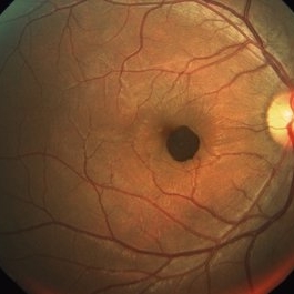
---thumb.jpg/image-square;max$300,300.ImageHandler) Benign Melanocytoma of the Optic Disc
Benign Melanocytoma of the Optic Disc
Jan 11 2013 by Hyung-Woo Kwak, MD
Fundus photography of optic disc showing a dark pigmented lesion.
Photographer: Dongho Kang, Kyung Hee Univsersity Hospital, Seoul
Imaging device: Zeiss f 450 plus
Condition/keywords: benign melanocytoma
-
 Central Chorioretinitis
Oct 3 2014 by Mehul A Shah
A 30-year-old female presented with complaint of sudden loss of vision OS before 2 months, on examination she was found to have pigmented lesion.
Photographer: Drashti Netralaya,Dahod
Imaging device: Zeiss ff450
-
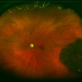
 Choroidal Melanocytosis
Sep 14 2023 by Ben Serar
Fundus photograph showing hyper pigmented lesion near the disc, at the level of the choroid, in a case of choroidal melanocytosis.
Condition/keywords: choroidal melanocytosis
-
 Choroidal Melanoma
Jan 29 2015 by H. Michael Lambert, MD
Dark pigmented lesion in retina with elevation.
-
 Choroidal naevus
Jan 11 2013 by Alex P. Hunyor, MD
Choroidal naevus with overlying drusen
Condition/keywords: benign pigmented lesions, choroidal nevus
-
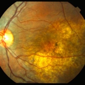 Choroidal Osteoma
Nov 21 2014 by Thomas A. Ciulla, MD, MBA, FASRS
This 13-year-old girl presented with mild painless progressive blurring of central vision left eye over the past several months. Visual acuity was 20/25. In the affected left eye, retinal examination revealed a relatively flat, lightly pigmented lesion, with well-defined and scalloped edges. Clumps of associated pigment were noted.
Photographer: Thomas Steele
Condition/keywords: choroidal neovascular membrane (CNVM), choroidal neovascularization (CNV), choroidal osteoma, macular choroidal osteoma
-
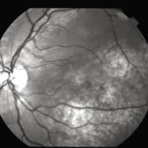
 Choroidal Osteoma
Nov 21 2014 by Thomas A. Ciulla, MD, MBA, FASRS
This 13-year-old girl presented with mild painless progressive blurring of central vision left eye over the past several months. Visual acuity was 20/25. In the affected left eye, retinal examination revealed a relatively flat, lightly pigmented lesion, with well-defined and scalloped edges. Clumps of associated pigment were noted. This OCT image shows subretiinal fluid just inferior to the fovea. Choroidal osteoma can be associated with the development of subretinal neovascularization (particularly at the edges of the osteoma).
Photographer: Thomas Steele
Condition/keywords: choroidal neovascular membrane (CNVM), choroidal neovascularization (CNV), choroidal osteoma, macular choroidal osteoma
-
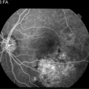
 Choroidal Osteoma
Nov 21 2014 by Thomas A. Ciulla, MD, MBA, FASRS
This 13-year-old girl presented with mild painless progressive blurring of central vision left eye over the past several months. Visual acuity was 20/25. In the affected left eye, retinal examination revealed a relatively flat, lightly pigmented lesion, with well-defined and scalloped edges. Clumps of associated pigment were noted. This OCT image shows subretiinal fluid just inferior to the fovea. Choroidal osteoma can be associated with the development of subretinal neovascularization (particularly at the edges of the osteoma).
Photographer: Thomas Steele
Condition/keywords: choroidal neovascular membrane (CNVM), choroidal neovascularization (CNV), choroidal osteoma, macular choroidal osteoma
-
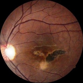
 CHRPE
CHRPE
Jan 15 2021 by Priya Rasipuram Chandrasekaran, MBBS, DO, DNB, FRCS
This is the fundus photo and fundus photo montage of the left eye of a 25-year-old male showing flat, solitary, round, greyish pigmented lesion situated AT THE equator with a scalloped margin. Vessels overlying the lesion are normal and there is a clear demarcation line between this and normal retina. The margins are hypopigmented with few hypopigmented lacunae inside.
Condition/keywords: congenital hypertrophy of the retinal pigment epithelium (CHRPE)
-
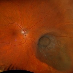 Collar Button Melanoma
Mar 27 2025 by Virginia Gebhart
62 year old male with large pigmented lesion with collar button. Pt states he was never aware of any lesion/nevus in the past. Fluid and orange pigment present, appears to be chronic. Pt will be scheduled for brachytherapy pending CT scan results.
Photographer: Virginia Gebhart, Retina Consultants of Carolina
Imaging device: Optos California
Condition/keywords: choroidal melanoma, collar button
-
 Congenital Hypertrophy of the Retinal Pigment Epithelium (CHRPE)
Aug 24 2012 by Andrew N. Antoszyk, MD FASRS
CHRPE lesion (black pigmented lesion) located along superior temporal arcade of left eye
Photographer: Lorainne Clark, Charlotte Eye Ear Nose and Throat Associates
-
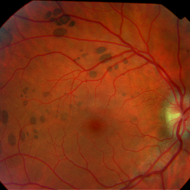
 Congenital Simple Hamartoma of the RPE Fundus Photo
Aug 3 2015 by Bindu Rajesh
Fundus photograph of a 26-year-old male ,showing a well defined pigmented lesion inferonasal to the fovea suggestive of simple hamartoma.
Imaging device: Visupac
Condition/keywords: congenital, hamartoma, retinal pigment epithelium
-
 Conjunctival Nevus
Dec 11 2014 by H. Michael Lambert, MD
Conjunctival Nevus- flat grey elevated pigmented lesion
Condition/keywords: nevus
-
---thumb.jpg/image-square;max$300,300.ImageHandler) Depigmented Lesion
Depigmented Lesion
Oct 29 2013 by Maurice F. Rabb
32 year old white female with depigmented lesion.
-
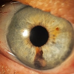 Iris Melanoma
Feb 1 2024 by Virginia Gebhart
90 year old female with elevated pigmented lesion, amelanotic portion extending toward the angle, questionable vascularity on UBM.
Photographer: Virginia Gebhart
Imaging device: Samsung Galaxy Z Flip
Condition/keywords: iris lesion, iris melanoma
 Loading…