 Initializing download.
-
 By Norman Byer
From Dr. Norman E. Byer’s “The Peripheral Retina in Profile” - Uploaded on Nov 9, 2012.
- Last modified by Suber S. Huang, MD, MBA, FASRS on Feb 11, 2013.
- Reviewed by Chayal Patel
- Rating
- Appears in
- Miscellaneous
- Condition/keywords
- scleral indentation, atrophic spot, pigmented lesion
- Description
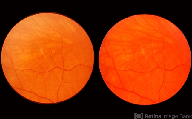
- The next three photographs are of the same lesion in a 26-year-old man and demonstrate the value of scleral indentation. This view without indentation shows only a tiny pigmented and atrophic spot in the fundus.


