Search results (1429 results)
-
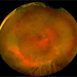
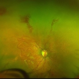
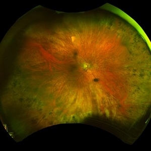
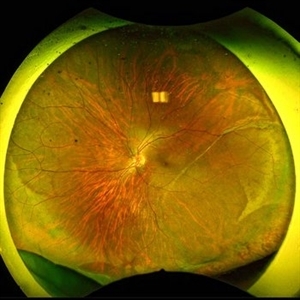 1 year Follow Up after Scleral Buckle Surgery in a Young Patient
May 18 2023 by Jesus Lozano, MD
25 year old man after Scleral Buckle Surgery + laser Retinopexy do to RRD macula off with ínfero temporal mid peripheral retinal holes in an area of lattice degeneration. Final VA 6/9.
Imaging device: Optos
Condition/keywords: scleral buckle
-
 5 Watt Blue Laser Pointer, Retinal Hemorrhage
Aug 30 2018 by John S. King, MD
5 watt laser pointer (class 4 laser pointer that can burn skin and material) to eye caused this hemorrhage as a result of injury to the retinal venule (see photo). Seen by Dr. Arnold, who sent this patient to Dr. Ware. Fortunately, fovea spared.
Imaging device: Optos
Condition/keywords: laser pointer retinopathy, retinal hemorrhage
-
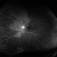
 Active Proliferative Diabetic Retinopathy
Jul 12 2024 by Korey Starkey
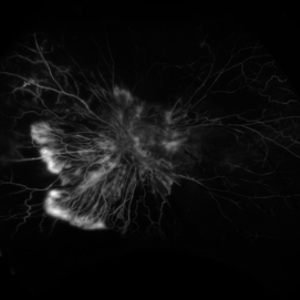
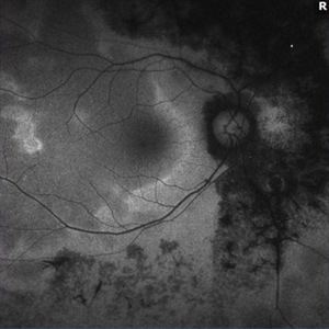
Fluorescein angiogram performed on 35 year old female with active proliferative diabetic retinopathy. Patient has peripapillary vascular loop and history of PRP treatment in both eyes. Patients left eye vision measured at Dcc20/200-1 at this visit.
Photographer: Korey Starkey
Imaging device: Optos
Condition/keywords: FLUORESCEIN ANGIOGRAPHY, hyperfluorescence, laser scarring, Optos, proliferative diabetic retinopathy (PDR), sea fan, ultra-wide field imaging, vascular loop
-
 Active Proliferative Diabetic Retinopathy
Aug 16 2022 by Donnie Willis
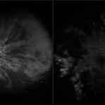
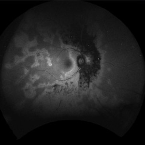
51 yo female. Uncontrolled Diabetes. Active Proliferative Diabetic Retinopathy.
Photographer: Donnie Willis, Tennessee Retina
Imaging device: Optos
Condition/keywords: capillary dropouts, Diabetes, fluorescein angiogram (FA), OPTOS, proliferative diabetic retinopathy (PDR), tractional retinal detachment
-
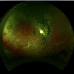
 Acute Central Retinal Artery Occlusion
Jul 27 2022 by Becca Harris
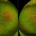
Ultra widefield FA/ICG of a 24 year old female with an acute central retinal artery occlusion affecting the right eye. Patient presented with extreme headaches following DAVF surgery the previous day. Patient has Factor VIII deficiency and had a cerebral venous thrombosis 9 years ago and lost vision in the right eye at that time. Patient has history of optic sheath fenestration OU and craniotomy. On initial evaluation, she had a CRAO as well as diffuse choroidal nonperfusion noted on optos FA. Suspect nonperfusion to third and sixth nerve leading to palsy. Occlusion of vasculature in the setting of recent endovascular embolization of fistulas in the CNS. Discussed diagnosis and poor prognosis with parents and patient. Patient had no light perception at the time of her initial appointment.
Photographer: Becca Harris
Imaging device: Optos California
Condition/keywords: Choroidal non-perfusion, fluorescein angiogram (FA), indocyanine green (ICG) angiography, non-perfusion, Optos, Right Eye, ultra-wide field imaging
-
 Acute Posterior Multifocal Placoid Pigment Epitheliopathy
Feb 20 2024 by Soobien Lee
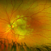
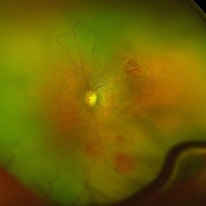
Optos color fundus photograph of a 20-year-old caucasian female with viral prodrome and vision loss OS>OD secondary to Acute Posterior Multifocal Placoid Pigment Epitheliopathy (APPME). Imaging of her left eye shows multiple bilateral creamy yellow-white placoid lesions at the level of RPE and choroid throughout the posterior pole.
Photographer: Ashley Metzger, Elman Retina Group
Imaging device: Optos Ultra-Widefield Imaging
Condition/keywords: acute posterior multifocal placoid pigment epitheliopathy (APMPPE), bacilliary layer detachment, Optos, uveitis, white dot syndrome
-
 Acute Posterior Multifocal Placoid Pigment Epitheliopathy
Feb 20 2024 by Soobien Lee
Optos fundus autofluorescence photograph of a 20-year-old caucasian female with viral prodrome and vision loss OS>OD secondary to Acute Posterior Multifocal Placoid Pigment Epitheliopathy (APPME). Imaging of her left eye shows hypoautofluorescent areas corresponding to multiple bilateral placoid lesions at the level of RPE and choroid throughout the posterior pole.
Photographer: Ashley Metzger, Elman Retina Group
Imaging device: Optos Ultra-Widefield Autoflurescence Imaging
Condition/keywords: acute posterior multifocal placoid pigment epitheliopathy (APMPPE), autofluorescence imaging, bacilliary layer detachment, Optos, OPTOS CALIFORNIA, uveitis, white dot syndrome
-
 Acute Posterior Multifocal Placoid Pigment Epitheliopathy
Feb 20 2024 by Soobien Lee
Fluorescein angiogram of a 20-year-old caucasian female with viral prodrome and vision loss OS>OD secondary to Acute Posterior Multifocal Placoid Pigment Epitheliopathy (APPME). Early blockage with late hyperfluorescent leakage can be seen on fluorescein angiography of the left eye.
Photographer: Ashley Metzger, Elman Retina Group
Imaging device: Optos Ultra-Widefield Fluorescein Angiography
Condition/keywords: acute posterior multifocal placoid pigment epitheliopathy (APMPPE), bacilliary layer detachment, FA, FA early phase, fluorescein angiogram (FA), Optos, uveitis, white dot syndrome
-
 Acute Posterior Multifocal Placoid Pigment Epitheliopathy
Feb 20 2024 by Soobien Lee
Fluorescein angiogram of a 20-year-old caucasian female with viral prodrome and vision loss OS>OD secondary to Acute Posterior Multifocal Placoid Pigment Epitheliopathy (APPME). Early blockage with late hyperfluorescent leakage can be seen on fluorescein angiography of the left eye.
Photographer: Ashley Metzger, Elman Retina Group
Imaging device: Optos Ultra-Widefield Fluorescein Angiography
Condition/keywords: acute posterior multifocal placoid pigment epitheliopathy (APMPPE), bacilliary layer detachment, FA, FA late phase, FA late phase leakage, fluorescein angiogram (FA), Optos, uveitis, white dot syndrome
-
 Acute Retinal Necrosis secondary to Herpes Zoster Ophthalmicus
Jan 9 2018 by Olivia Rainey
Ultra-wide field Optos pseudocolor montage of an 40-year-old female presenting with acute retinal necrosis secondary to herpes zoster ophthalmicus affecting her right eye.
Photographer: Olivia Rainey
Imaging device: Optos California
Condition/keywords: acute retinal necrosis, color fundus photograph, Herpes zoster, montage, Optos, ultra-wide field imaging
-
 Acute Syphilitic Posterior Placoid Chorioretinitis
Nov 22 2020 by Shawn Sell
58-year-old homeless male presenting with 2 weeks of bilateral eye redness and photosensitivity found to have panuveitis with a positive VDRL CSF and RPR titer of 1:512 with acute syphilitic posterior placoid chorioretinitis.
Photographer: Eastern Virginia Medical School
Imaging device: Optos
Condition/keywords: acute syphilitic posterior placoid chorioretinitis
-
 Acute Syphilitic Posterior Placoid Chorioretinitis
Nov 22 2020 by Shawn Sell
58-year-old homeless male presenting with 2 weeks of bilateral eye redness and photosensitivity found to have panuveitis with a positive VDRL CSF and RPR titer of 1:512 with acute syphilitic posterior placoid chorioretinitis.
Photographer: Eastern Virginia Medical School
Imaging device: Optos
Condition/keywords: acute syphilitic posterior placoid chorioretinitis, neurosyphilis
-
 Acute Zonal Occult Outer Retinopathy
Dec 16 2020 by Robert C Wann, MD
Fundus photo of a 28-year-old female with AZOOR.
Photographer: Retina Consultants of Alabama
Imaging device: Optos
Condition/keywords: acute zonal occult outer retinopathy (AZOOR)
-
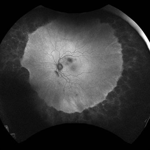
 Acute Zonal Occult Outer Retinopathy
Dec 16 2020 by Robert C Wann, MD
Fundus autofluorescence of a 28-year-old female with AZOOR.
Photographer: Retina Consultants of Alabama
Imaging device: Optos
Condition/keywords: acute zonal occult outer retinopathy (AZOOR)
-
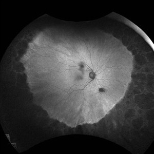
 Acute Zonal Occult Outer Retinopathy
Dec 16 2020 by Robert C Wann, MD
Fundus autofluorescence of a 28-year-old female with AZOOR.
Photographer: Retina Consultants of Alabama
Imaging device: Optos
Condition/keywords: acute zonal occult outer retinopathy (AZOOR)
-
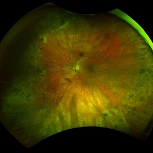
 Acute Zonal Occult Outer Retinopathy (AZOOR)
Jan 19 2022 by James B. Soque, CRA, OCT-C, COA, FOPS
Acute Zonal Occult Outer Retinopathy, OD. Color fundus, Ultra-wide field photograph. 46-year-old white male, VA CC 10/16, 20/12.5, has had recurrent vasculitis for 11 years. No treatment.
Photographer: James Soque, CRA, OCT-C, COA, FOPS, Island Retina, Shirley, NY
Imaging device: Optos California
Condition/keywords: acute zonal occult outer retinopathy (AZOOR), color wide field, optos, ultra-wide field imaging
-
 Adult Onset Coats' Disease
Jul 5 2024 by Zach Seim
FA/ICG of a 64 year old female with Adult Onset Coats' Disease. VA DCC CF@3 feet upon presentation. Therapy options discussed extensively.
Photographer: Zach Seim
Imaging device: Optos California
Condition/keywords: Coats' disease, FA, indocyanine green (ICG) angiography, Optos, OPTOS CALIFORNIA
-
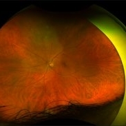
 Advanced RP
Nov 5 2024 by rahul saradge
A man, 58, arrived complaining of BOV for both near and distance vision in both eyes, with a 6/9 BCVA in each eye. For a year, the patient had been taking medication for both diabetes and hypertension. In both eyes, the dilated ophthalmoscopic retina revealed waxy disc pallor paired with bony spicules in the mid-periphery. The patient was prescribed spectacles and given counseling regarding the nature of the illness.
Photographer: Lokesh Dukare ,Isha Netralaya Thane
Imaging device: optos
Condition/keywords: bone spicule, optic disc pallor, Optos, Retinitis Pigmentosa
-
 Aggressive ROP
Oct 26 2024 by rahul saradge
Premature baby referred for ROP evaluation. HALF ZONE 1 was only vascularised, Patient was given Inj Anti-Vegf followed by ROP Laser after 1 week.
Photographer: Ankita Choudhary ,Isha Netralaya Thane
Imaging device: optos
Condition/keywords: aggresive retinopathy of prematurity, optos, retinopathy of prematurity
-
 Aggressive ROP
Oct 26 2024 by rahul saradge
Premature baby referred for ROP evaluation. HALF ZONE 1 was only vascularised, Patient was given Inj Anti-Vegf follwed by ROP Laser after 1 week
Photographer: Neha Choudhary,Isha Netralaya Thane
Imaging device: optos
Condition/keywords: aggressive posterior retinopathy of prematurity (APROP), retinopathy of prematurity
-
 Alagille Syndrome UWF Autofluorescence
Dec 4 2023 by Isaac Ezon, MD
43 yo Female with known Alagille Syndrome, referred for peripheral retinal changes. Subjective nyctalopia but no other symtpoms. Alagille Syndrome UWF Autofluorescence.
Photographer: Tara Murray
Imaging device: Optos
Condition/keywords: hereditary choroidal dystrophy, hereditary retinal degeneration
-
 Alagille Syndrome UWF Color
Dec 4 2023 by Isaac Ezon, MD
43 yo Female with known Alagille Syndrome, referred for peripheral retinal changes. Subjective nyctalopia but no other symtpoms. Alagille Syndrome UWF Color.
Photographer: Tara Murray
Imaging device: Optos
Condition/keywords: hereditary choroidal dystrophy, hereditary retinal degeneration
-
 Amelanotic Choroidal Melanoma
Apr 12 2019 by David L Kilpatrick, MD
Fundus photograph of a 69-year-old male with an amelanotic choroidal melanoma and corresponding exudative retinal detachment. Transvitreal biopsy was performed at the time of radioactive I-125 plaque placement. The genetic expression profile revealed a Class 1A, PRAME negative tumor.
Photographer: Retina Consultants of Alabama, P. C.
Imaging device: Optos
Condition/keywords: amelanotic melanoma
 Loading…