Search results (67 results)
-
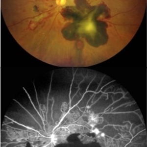
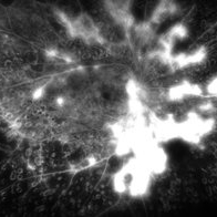 Active neovascularization in Proliferative Diabetic Retinopathy
Jan 10 2018 by Peter H. Tang, MD, PhD
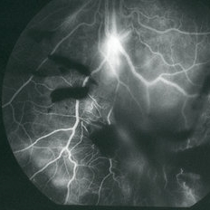
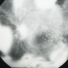
Fluorescein angiography image from a 46-year-old woman with uncontrolled proliferative diabetic retinopathy shows extensive dye leakage from active neovascularization.
Imaging device: Optos California
Condition/keywords: diabetes, diabetic retinopathy, fluorescein leakage, neovascularization elsewhere (NVE), neovascularization of the disc (NVD), pan-retinal photocoagulation (PRP), proliferative diabetic retinopathy (PDR)
-
 BRVO With Laser
Feb 19 2015 by H. Michael Lambert, MD
Color photo of NVE after BRVO. Laser performed, photos just after laser and a few weeks later.
Condition/keywords: branch retinal vein occlusion (BRVO), laser, neovascularization elsewhere (NVE)
-
 BRVO With PRP laser
Feb 19 2015 by H. Michael Lambert, MD
Color photo of NVE after BRVO. Laser performed,ERM present
Condition/keywords: ischemia, neovascularization elsewhere (NVE)
-
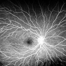 Coats' Disease
Oct 20 2020 by Anfisa Ayalon, MD
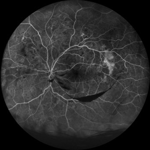
Fundus fluorescein angiography of 35-year-old female with right eye asymptomatic coats disease.
Photographer: Anfisa Ayalon, Meir Medical Center, Kfar Saba, Israel.
Imaging device: California, Optos 200 DTX
Condition/keywords: Coats' disease, neovascularization elsewhere (NVE), retina
-
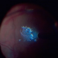 Detached NVE During PVD induction
Apr 27 2018 by Michael J. Koss, MD, PhD, MBA
A 73-year-old woman with macular pucker underwent a pars plana vitrectomy with membrane peeling. Additionally the patient suffers from diabetic retinopathy after being diagnosed with type 2 diabetes mellitus sixteen years ago. Prior to the procedure she was treated with a series of intravitreal Bevacizumab-injections due to diabetic macular edema. There was no history of a proliferative DRP. During the vitrectomy a branch of an obliterated NVE spontaneously detached and floated freely in the vitreous. The 3D shot was captured via Alcon’s NGENUITY® 3D Visualization System in form of photograph and video providing an outstandingly detailed image of the branched NVE.
Photographer: Michael Koss, Augenzentrum Nymphenburger Hoefe
Imaging device: Alcon’s NGENUITY® 3D Visualization System
Condition/keywords: diabetes, diabetic retinopathy, neovascularization elsewhere (NVE), pars plana vitrectomy (PPV), PVD induction
-
 Diabetic Tractional Retinal Detachment
Jan 10 2018 by Peter H. Tang, MD, PhD
Fundus photograph of a 46-year-old woman with proliferative diabetic retinopathy and tractional retinal detachment that is poorly controlled.
Imaging device: Optos California
Condition/keywords: diabetes, neovascularization elsewhere (NVE), neovascularization of the disc (NVD), pan-retinal photocoagulation (PRP), proliferative diabetic retinopathy (PDR), retinal fibrosis, tractional retinal detachment
-
 Eales Disease
Apr 1 2019 by Gary R. Cook, MD, FACS
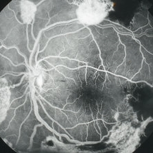
Late-phase (5 minutes) fluorescein angiogram image of the nasal mid-periphery of the left eye of a 23-year-old Vietnamese female with Eales Disease showing multiple areas of NVE and some disc leakage.
Imaging device: Topcon VT-50
Condition/keywords: Eales disease, FA late phase, FA late phase leakage, fluorescein angiogram (FA), neovascularization elsewhere (NVE)
-
 Eales Disease
Apr 1 2019 by Gary R. Cook, MD, FACS
Mid-phase (70 seconds) fluorescein angiogram image of the inferior periphery OS of a 20-year-old Vietnamese male with Eales Disease; there is bright hyperfluorescence from a focus of NVE below the optic disc and blocked fluorescence from vitreous hemorrhage in the eye.
Imaging device: Topcon VT-50
Condition/keywords: Eales disease, FA late phase, FA late phase leakage, fluorescein angiogram (FA), neovascularization elsewhere (NVE), vitreous hemorrhage
-
 Eales Disease
Apr 1 2019 by Gary R. Cook, MD, FACS
Late-phase (5 minutes) fluorescein angiogram image of a 20-year-old Vietnamese male with Eales Disease showing retinal vascular changes and intense leakage from peripheral NVE.
Imaging device: Topcon VT-50
Condition/keywords: Eales disease, FA late phase leakage, fluorescein angiogram (FA), neovascularization elsewhere (NVE)
-
 Eales Disease
Apr 1 2019 by Gary R. Cook, MD, FACS
Mid-phase fluorescein angiogram image of the left eye of a 23-year-old Vietnamese female with Eales Disease showing the retinal vascular abnormalities, capillary loss, and a focus of NVE; V.A.= 20/25-2.
Imaging device: Topcon VT-50
Condition/keywords: Eales disease, FA mid phase, fluorescein angiogram (FA), neovascularization elsewhere (NVE)
-
 Eales Disease
Apr 1 2019 by Gary R. Cook, MD, FACS
Mid-phase fluorescein angiogram frame of the left eye of a 23-year-old Vietnamese female with Eales Disease showing multiple areas of NVE and areas of capillary loss and nonperfusion OS.
Imaging device: Topcon VT-50
Condition/keywords: Eales disease, FA mid phase, fluorescein angiogram (FA), neovascularization elsewhere (NVE)
-
 Eales Disease
Apr 1 2019 by Gary R. Cook, MD, FACS
Late-phase fluorescein angiogram image of the left eye of a 23-year-old Vietnamese female with Eales Disease showing extensive dye leakage from multiple areas of NVE and from some NVD.
Imaging device: Topcon VT-50
Condition/keywords: Eales disease, FA late phase, fluorescein angiogram (FA), neovascularization elsewhere (NVE)
-
 Fluorescein Angiography Neovascularization Elsewhere and Subhyaloid Hemorrhage
Aug 15 2021 by ASRS Staff
38 year-old male, presented with complaint of dark spot in vision of left eye. His vision was 6/6 in both eyes. On examination he was having subhyaloid hemorrhage and NVE in left eye.NVE was also present in RE. Patient was referred for carotid Doppler and cardiologist opinion.
Imaging device: Nidek Mirante
Condition/keywords: neovascularization elsewhere (NVE), subhyaloid hemorrhage
-
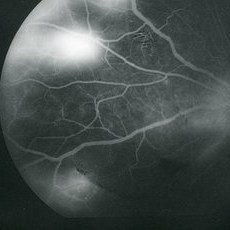
 High-Risk Proliferative Diabetic Retinopathy
Mar 20 2019 by Anfisa Ayalon, MD
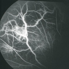
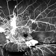
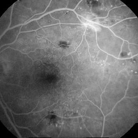
Fundus fluorescein angiography of 58-year-old patient with left eye high-risk proliferative diabetic retinopathy. Note severe ischemia of retina, large areas of neovascularization elsewhere and preretinal hemorrhages.
Photographer: Anfisa Ayalon,MD., Meir Medical Center, Kfar Saba, Israel.
Imaging device: California, Optos 200 DTX
Condition/keywords: ischemia, neovascularization elsewhere (NVE), proliferative diabetic retinopathy (PDR), retina, subhyaloid hemorrhage
-
 Hypertensive Retinopathy
Dec 24 2017 by Purva Patwari
52-year-old female diagnosed of hypertension by retina evaluation.
Photographer: Dr Purva Patwari, Patwari Retina Center, Ahmedabad, Gujarat , India
Imaging device: ZEISS VISU500
Condition/keywords: hypertensive retinopathy, neovascularization elsewhere (NVE), Roth spots
-
 Lightening In Eyes
Jul 22 2021 by Vishal Gupta, MBBS, MS
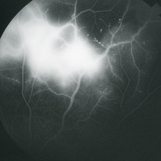
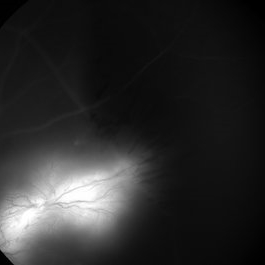
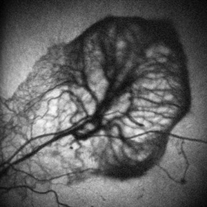
Fundus fluorescein angiogram of a neovascular frond lighting up like a silent lightening thunder in the dark night.
Photographer: Dr Vishal Gupta, INHS Asvini, Mumbai, INDIA
Imaging device: zeiss
Condition/keywords: branch retinal vein occlusion (BRVO), fluorescein angiogram (FA), neovascularization elsewhere (NVE)
-
 Lizard-shaped-hemorrhage
Apr 29 2023 by Saagar A Pandit, MD, MPH
49 year-old male with a history of Type 1 Diabetes Mellitus, past ocular history significant for proliferative diabetic retinopathy of both eyes. Left eye significant for a unique, "lizard-shaped" pre-retinal hemorrhage in an area of neovascularization. Note the corresponding fluorescein angiography which demonstrates blockage from hemorrhage and significant posterior non-perfusion, in addition to tufts of neovascularization which are hyperfluorescent.
Photographer: Maria Pei, Bellevue Hospital Ophthalmology Clinic, New York, NY
Condition/keywords: hemorrhage, neovascularization elsewhere (NVE), proliferative diabetic retinopathy (PDR)
-
 Neovascularization - RDP
Jun 29 2014 by Ratimir Lazic, MD, PhD
A FAG image of a 84-year-old female. Late venous phase of the left eye. Hyperfloercent area in upper temporal quadrant represents NVE. Many hyperflorescent dots can be seen. Few hypoflorescent areas are deep retinal hemorrhages.
Photographer: Marko Lukic, University Eye Clinic Svjetlost
Imaging device: Zeis Visucam Lite 2
Condition/keywords: neovascularization (NV), neovascularization elsewhere (NVE)
-
 Neovascularization - RDP
Jun 29 2014 by Ratimir Lazic, MD, PhD
FAG image of a 84-year-old female. Dye leakage from the NVE can be seen.
Photographer: Marko Lukic, University Eye Clinic Svjetlost
Imaging device: Zeis Visucam Lite 2
Condition/keywords: neovascularization elsewhere (NVE)
-
 Neovascularization Elsewhere
Jan 16 2018 by Jay G. Prensky, MD
Example of fluorescein angiogram of neovascularization at border of treated and untreated nonperfused retina in patient with proliferative diabetic retinopathy
Imaging device: Optos
Condition/keywords: neovascularization elsewhere (NVE), pan-retinal photocoagulation (PRP), proliferative diabetic retinopathy (PDR)
-
 Neovascularization Elsewhere in Proliferative Diabetic Retinopathy
Nov 16 2019 by Anfisa Ayalon, MD
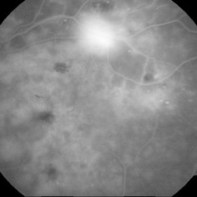
Fundus autofluorescence image of neovascularization elsewhere, patient with high-risk proliferative diabetic retinopathy.
Photographer: Anfisa Ayalon, MD., Meir Medical Center, Kfar Saba, Israel.
Condition/keywords: diabetes, fundus autofluorescence (FAF), ischemia, neovascularization elsewhere (NVE), proliferative diabetic retinopathy (PDR)
-
 NVE from BRVO
Feb 19 2015 by H. Michael Lambert, MD
Color photo of NVE after BRVO.
Condition/keywords: branch retinal vein occlusion (BRVO), neovascularization elsewhere (NVE)
-
 NVE from BRVO
Feb 19 2015 by H. Michael Lambert, MD
Color photo of NVE after BRVO.
Condition/keywords: branch retinal vein occlusion (BRVO), neovascularization elsewhere (NVE)
-
 NVE from BRVO
Feb 19 2015 by H. Michael Lambert, MD
Color photo of NVE after BRVO.
Condition/keywords: branch retinal vein occlusion (BRVO), neovascularization elsewhere (NVE)
-
 NVE in a Patient With Vasculitis
Nov 5 2018 by awaneesh m upadhyay, MBBS, DNB
FFA image of a 22-year-old male vasculitis patient with NVE.
Photographer: Hiteshwar Saikia
Condition/keywords: neovascularization elsewhere (NVE), tuberculosis, vasculitis
 Loading…