Search results (150 results)
-
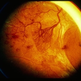 Advanced Active PDR
Mar 29 2013 by Henry J. Kaplan, MD
Large active NVEs with fibrous proliferations in diabetes.
Condition/keywords: fibrous proliferation, neovascularization (NV)
-
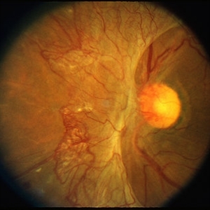 Advanced Active PDR
Mar 29 2013 by Henry J. Kaplan, MD
Extensive NVD-FPD and NVE-FPE in a diabetic patient.
Condition/keywords: foveal photoreceptor defect, FPE, neovascularization (NV), neovascularization of the disc (NVD)
-
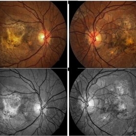 Angioid Streaks
May 11 2016 by Andrea Arriola-Lopez, MD MSc
64-year-old man, VA CF AO. Inactive neovascularization. Color fundus and red free photograph.
Photographer: Andrea E. Arriola-Lopez MD MSc
Imaging device: Visucam lite Zeiss
Condition/keywords: angioid streaks, color fundus photograph, neovascularization (NV), red-free
-
---thumb.jpg/image-square;max$300,300.ImageHandler) Anterior Hyaloid Fibrovascular Proliferation
Anterior Hyaloid Fibrovascular Proliferation
Feb 13 2013 by From the Collections of Thomas M. Aaberg, MD and Thomas M. Aaberg Jr., MD
Histopathology neovascularization.
Condition/keywords: fibrovascular proliferation, histopathology, hyaloid, neovascularization (NV)
-
 Battle of BRVOs: Old vs New
Jul 30 2021 by Gayathri Mohan
Color fundus photograph of a 48-year-old showing an inferno temporal BRVO. Old BRVO with neovascularization seen super-temporally.
Photographer: Dr. Gayathri Mohan
Imaging device: Canon
Condition/keywords: branch retinal vein occlusion (BRVO), cystoid macular edema (CME), neovascularization (NV)
-
 Bilateral Retinal Vaso-Occlusive Disease
Jun 20 2017 by S. Natarajan, MD, FASRS, FRCS (GLASGOW) , FICO, D.Sc, FELA
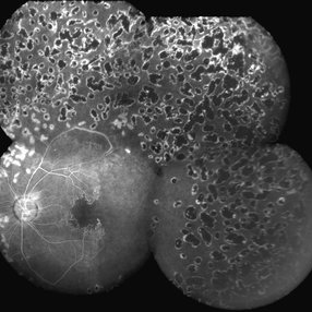
35-year-old woman with bilateral retinal vaso-occlusive disease with secondary neovascularization. She has undergone extensive care including photocoagulation to both the retina. There is severe ischemia on the posterior pole in both eyes. Patient has been started on immunosuppressant.
Photographer: Ms . Ashwini Borde
Imaging device: zeiss plus IR 450
Condition/keywords: neovascularization (NV), vaso-occlusive disease
-
 Branch Retinal Artery Occlusion
Sep 11 2018 by Olivia Rainey
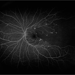
Ultra-wide field fluorescein angiogram of a 46-year-old male with a branch retinal artery occlusion affecting his left eye. The longstanding occlusion and has resulted in peripheral nonperfusion and neovascularization.
Photographer: Olivia Rainey
Imaging device: Optos
Condition/keywords: branch retinal artery occlusion (BRAO), fluorescein angiogram (FA), left eye, neovascularization (NV), non-perfusion, Optos
-
 Branch Retinal Vein Occlusion
Sep 28 2017 by Purva Patwari
Patient came for routine work.
Photographer: Dr Purva Patwari,Patwari Retina Clinic,Ahmedabad
Imaging device: Zeiss
Condition/keywords: neovascularization (NV)
-
 Branch Vein Occlusion (BVO) (NV)
Oct 1 2013 by Howard Schatz, MD
Fifty year old female, right eye 20/30, left eye 20/40.
Condition/keywords: branch vein occlusion (BVO), neovascularization (NV)
-
 Branch Vein Occlusion (BVO) (NV)
Oct 1 2013 by Howard Schatz, MD
Fifty six year old white female, right eye 5/200, left eye 20/15.
Condition/keywords: branch vein occlusion (BVO), neovascularization (NV)
-
 Branch Vein Occlusion (BVO) (NV)
Oct 1 2013 by Howard Schatz, MD
Sixty five year old white female, right eye 20/15, left eye 20/50.
Condition/keywords: branch vein occlusion (BVO), neovascularization (NV)
-
 Branch Vein Occlusion (BVO) (NV)
Oct 1 2013 by Howard Schatz, MD
Fifty one year old female, right eye 20/40, left eye 20/30.
Condition/keywords: branch vein occlusion (BVO), neovascularization (NV)
-
 Branch Vein Occlusion (BVO) (NV)
Oct 1 2013 by Howard Schatz, MD
Sixty year old white male, right eye 20/250, left eye 20/20.
Condition/keywords: branch vein occlusion (BVO), neovascularization (NV)
-
 Branch Vein Occlusion (BVO)(NV)
Oct 1 2013 by Howard Schatz, MD
Sixty eight year old male, right eye 20/40, left eye 20/20.
Condition/keywords: branch vein occlusion (BVO), neovascularization (NV)
-
 BRVO Complications
Mar 29 2013 by Henry J. Kaplan, MD
Old superotemporal BRVO as a sclerotic vessel with NVD and NVE and vitreous hemorrhage and a preretinal hemorrhage.
Condition/keywords: branch retinal vein occlusion (BRVO), neovascularization (NV), neovascularization of the disc (NVD), vitreous hemorrhage
-
---thumb.jpg/image-square;max$300,300.ImageHandler) Cartoon Subretinal NV
Cartoon Subretinal NV
Feb 13 2013 by From the Collections of Thomas M. Aaberg, MD and Thomas M. Aaberg Jr., MD
Cartoon subretinal NV.
Condition/keywords: cartoon, neovascularization (NV), subretinal
-
 Cornea
Feb 23 2018 by JEFFERSON R SOUSA, Tecg.º (Biomedical Systems Technology)
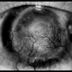
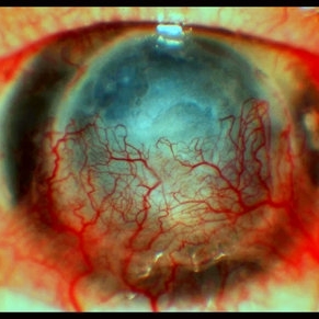
64-year-old, patient with vision loss more than 10 years after having suffered blunt trauma with ocular perforation.
Photographer: JEFFERSON R SOUSA - Study Center and Ophthalmological Research Dr. Andre M V Gomes, Institute Dr. Suel Abujamra São Paulo-Brazil
Imaging device: Topcon TRC-50 DX, Imaginet 5.0, angle de 20 graus. Flash 36.
Condition/keywords: 20 degrees, central opacity of cornea, corneal edema, neovascularization (NV)
-
 CORNEA
Feb 23 2018 by JEFFERSON R SOUSA, Tecg.º (Biomedical Systems Technology)
64-year-old patient, with vision loss more than 10 years after having suffered blunt trauma with ocular perforation.
Photographer: JEFFERSON R SOUSA - Study Center and Ophthalmological Research Dr. Andre M V Gomes, Institute Dr. Suel Abujamra São Paulo-Brazil
Imaging device: Topcon TRC-50 DX, Imaginet 5.0, angle de 20 graus. Flash 36.
Condition/keywords: 20 degrees, central opacity of cornea, corneal edema, neovascularization (NV)
-
 Diabetic Macular Edema, Proliferative Diabetic Retinopathy, Neovascularization Elsewhere, DME, PDR, NVE
Apr 1 2013 by James B. Soque, CRA, OCT-C, COA, FOPS
39-year-old white female and long standing diabetis, c/o new peripheral symptoms of left eye. FA OS reveals diabetic macular edema, microaneurysms, and neovasculaization elsewhere. Fluorescein Angogram, Early Phase, 50 Deg, 2x Mag.
Photographer: James B Soque, CRA, COA
Imaging device: Topcon TRC 50DX with MERGE software, OIS 10.6.45
Condition/keywords: diabetic macular edema, neovascularization (NV), proliferative diabetic retinopathy (PDR)
-
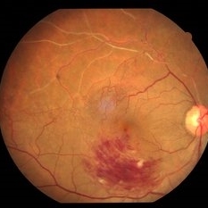
Diabetic Neovascularization
Oct 8 2012 by David R. Chow, MD, FRCS(C)
Diabetic neovascularization.
Condition/keywords: neovascularization (NV)
-
 Diabetic Proliferative Retinopathy
Dec 1 2019 by Lucas Zago Ribeiro, MD
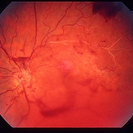
Fundus photograph of 75-year-old man with diabetic proliferative retinopathy with fibrovascular proliferation over the optic disc.
Photographer: Lucas Zago Ribeiro, Federal University of São Paulo
Imaging device: Zeiss Visucam 524
Condition/keywords: diabetic retinopathy, fibrovascular proliferation, neovascularization (NV)
-
 Diabetic Retinopathy
Aug 21 2015 by Andrea Arriola-Lopez, MD MSc
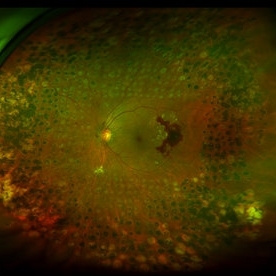
Color fundus photography shows neovascularization of the optic nerve head, macular pre retinal hemorrhage, pan retinal photocoagulation and extreme temporal peripherical retina without PRP.
Photographer: Andrea Elizabeth Arriola L.
Imaging device: OPTOS Dakota
Condition/keywords: diabetes, diabetic retinopathy, neovascularization (NV)
-
 Diabetic Traction Detachment of Retina
Sep 28 2022 by Chloe Hanifan
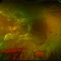
Ultra-widefield pseudo color fundus photograph of a 50-year-old female with a Diabetic Traction Detachment of Retina affecting her left eye. The patient was unable to proceed with surgery due to other health issues in May of 2022, when she presented in the office in September of 2022, a guarded prognosis given chronicity and associated ischemia. The patient was LP at the time of the September appointment.
Photographer: Chloe Hanifan
Imaging device: Optos California
Condition/keywords: diabetes, diabetic traction detachment, fundus photography, left eye, neovascularization (NV), Optos, proliferative diabetic retinopathy (PDR), pseudocolor, ULTRA WIDE FIELD
-
 Diabetic Traction Detachment of Retina
Sep 28 2022 by Chloe Hanifan
Ultra-widefield pseudo color fundus photograph of a 50-year-old female with a Diabetic Traction Detachment of Retina affecting her left eye. The patient was unable to proceed with surgery due to other health issues in May of 2022, when she presented in the office in September of 2022, a guarded prognosis given chronicity and associated ischemia. The patient was LP at the time of the September appointment.
Photographer: Chloe Hanifan
Imaging device: Optos California
Condition/keywords: Diabetes, diabetic traction detachment, fundus photography, left eye, neovascularization (NV), Optos, proliferative diabetic retinopathy (PDR), pseudocolor, ULTRA WIDE FIELD
-
 Extensive Neovascularization
Jul 21 2024 by César Adrián Gómez Valdivia, MD
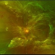
Macular Traction and Extensive Neovascularization found in a 66 year-old patient with history of uncontrolled Type 2 Diabetes Mellitus.
Photographer: Erika Paulina Ornelas Cazares
Imaging device: TOPCON TRC-50DX
Condition/keywords: diabetic retinopathy, neovascularization (NV)
 Loading…