Search results (3038 results)
-
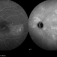
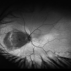
 Acute Macular Neuroretinopathy
Dec 11 2019 by Lauren Whaley
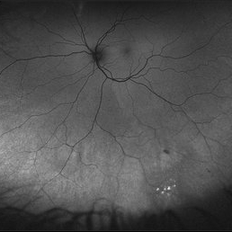
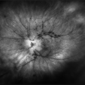
34-year-old female patient presented with changes in vision after recent upper respiratory infection. Referring doctor originally thought it was a blood pressure issue. She noticed a "C" shape in her vision. Infrared image was captured showing exactly what patient was describing! Doctor confirmed with this image that it was AMN.
Photographer: Lauren R. Whaley, COA
Imaging device: Heidelberg Spectralis
Condition/keywords: 30 degrees, acute macular neuroretinopathy, Heidelburg Spectralis, left eye, macula, near infrared autofluorescence (NIRAF)
-
 Acute Retinal Necrosis
Oct 10 2012 by Jeffrey G. Gross, MD, FASRS
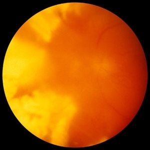
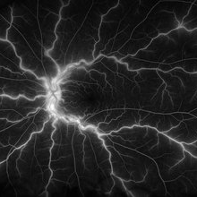
Acute retinal necrosis, left eye.
Condition/keywords: acute retinal necrosis, left eye
-
---thumb.jpg/image-square;max$300,300.ImageHandler) Adult Vitelliform Dystrophy
Adult Vitelliform Dystrophy
Feb 13 2013 by From the Collections of Thomas M. Aaberg, MD and Thomas M. Aaberg Jr., MD
Left eye
Condition/keywords: left eye, vitelliform macular dystrophy
-
 Autofluorescence of Peripheral Retinoschisis
Jul 26 2018 by Olivia Rainey
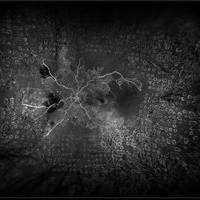
Ultra-wide field autofluorescence image of a 49-year-old male with non-progressive peripheral retinoschisis of his left eye. Patient was asymptomatic and had no prior trauma or surgery to his eye. Recommended observation at this time.
Photographer: Olivia Rainey
Imaging device: Optos
Condition/keywords: autofluorescence imaging, left eye, Optos, retinoschisis, ultra-wide field imaging
-
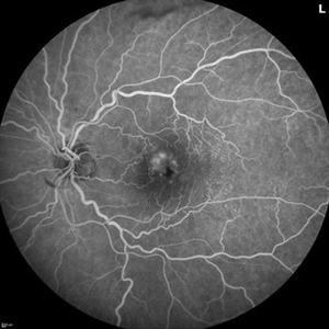
 Birdshot Chorioretinopathy FA Mid Phase Left Eye
Oct 9 2012 by Jeffrey G. Gross, MD, FASRS
Birdshot chorioretinopathy, FA, mid phase, left eye.
Condition/keywords: birdshot, chorioretinopathy, left eye, mid phase
-
 Birdshot Chorioretinopathy FA with Optic Nerve Leakage Left Eye
Oct 9 2012 by Jeffrey G. Gross, MD, FASRS
Birdshot chorioretinopathy, FA, with optic nerve leakage, left eye.
Condition/keywords: birdshot, chorioretinopathy, left eye, optic nerve leakage
-
 Birdshot Chorioretinopathy FA with Optic Nerve Leakage Left Eye
Oct 9 2012 by Jeffrey G. Gross, MD, FASRS
Birdshot chorioretinopathy, FA, with optic nerve leakage, left eye.
Condition/keywords: birdshot, chorioretinopathy, left eye, optic nerve leakage
-
 Birdshot Chorioretinopathy FA with Patchy Choroidal Filling Left Eye
Oct 9 2012 by Jeffrey G. Gross, MD, FASRS
Birdshot chorioretinopathy, FA, with patchy choroidal filling, left eye.
Condition/keywords: birdshot, chorioretinopathy, left eye, patchy choroidal filling
-
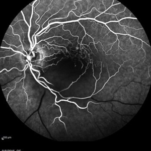
 Branch Retinal Artery Occlusion
Sep 11 2018 by Olivia Rainey
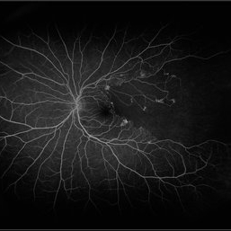
Ultra-wide field fluorescein angiogram of a 46-year-old male with a branch retinal artery occlusion affecting his left eye. The longstanding occlusion and has resulted in peripheral nonperfusion and neovascularization.
Photographer: Olivia Rainey
Imaging device: Optos
Condition/keywords: branch retinal artery occlusion (BRAO), fluorescein angiogram (FA), left eye, neovascularization (NV), non-perfusion, Optos
-
 Branch Retinal Vein Occlusion
Sep 11 2018 by Olivia Rainey
Ultra-wide field pseudocolor montage of an 84-year-old female with a branch retinal vein occlusion affecting her left eye. Patient recently had a PPV for a epiretinal membrane in her left eye and shortly after developed an occlusion.
Photographer: Olivia Rainey
Imaging device: Optos
Condition/keywords: branch retinal artery occlusion (BRAO), hemorrhage, left eye, montage, Optos, pseudocolor, ultra-wide field imaging
-
 Branch Retinal Vein Occlusion with Multifactorial Macular Edema and Epiretinal Membrane
Oct 3 2024 by Logan ryzenga
Fluorescein angiogram of a 62 year old woman with cystoid macular edema from concurrent Epiretinal Membrane and Branch Retinal Vein occlusion. She has an extensive history of anti-VEGF injections with stable but unresolved macular edema. Following angiography, it was determined that an epiretinal membrane peel would be indicated in an attempt to achieve resolution of macular edema.
Photographer: Logan Ryzenga
Imaging device: Heidelberg Spectralis
Condition/keywords: 55-degrees, branch retinal vein occlusion (BRVO), cystoid macular edema (CME), epiretinal membrane (ERM), Fluorescein angiography, heidelberg spectralis, hyperfluorescence, leakage, left eye, OS, wide angle imaging
-
 Branch Retinal Vein Occlusion With Peripheral Pigmentary Change
Jan 15 2019 by Olivia Rainey
Ultra-wide field fluorescein angiogram of an 85-year-old female with a branch retinal vein occlusion with peripheral pigmentary changes. Patient developed a BRVO after a PPV for an epiretinal membrane.
Photographer: Olivia Rainey
Imaging device: Optos
Condition/keywords: branch retinal vein occlusion (BRVO), epiretinal membrane (ERM), fluorescein angiogram (FA), left eye, Optos, pigmentary retinal dystrophy
-
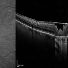 Bullseye Maculopathy
Jan 22 2024 by Kali Jend
Optical coherence tomography of a 73-year-old female with Bullseye Macular Changes affecting her left eye. Patient reports having a family history of this condition and denies prior Plaquenil or Elmiron use. Compared to previous imaging, the patient's condition progressed in the left eye from 2020 to 2023. Patient has a history of fluctuating Diabetic Macular Edema and a current Epiretinal Membrane as well. Patient's vision was Ncc20/60 at the time the image was taken.
Photographer: Kali Jend
Imaging device: Heidelberg Spectralis
Condition/keywords: bullseye maculopathy, epiretinal membrane (ERM), heidelberg spectralis, left eye, macular pucker, OCT, optical coherence tomography (OCT)
-
 Central Retinal Artery Occlusion
May 16 2017 by Olivia Rainey
Fluorescein angiogram of an 66-year-old female with a central retinal artery occlusion affecting her left eye.
Photographer: Olivia Rainey
Imaging device: Heidelberg Spectralis
Condition/keywords: 50 degrees, central retinal artery occlusion (CRAO), fluorescein angiogram (FA), left eye, mid phase, retinal ischemia
-
 Central Retinal Artery Occlusion Secondary to Endophthalmitis
Oct 24 2022 by Kelli Nyenhuis
Ultra-widefield fluorescein angiogram of a 64 year old female who developed a Central Retinal Artery Occlusion following acute endophthalmitis. The physician commented that there is no vascular filling with the exception of the papillomacular bundle. The patient's vision was scHM at the time the image was taken.
Photographer: Kelli Nyenhuis
Imaging device: Optos California
Condition/keywords: central retinal artery occlusion (CRAO), endophthalmitis, fluorescein angiogram (FA), left eye, non-perfusion, Optos, ultra-wide field imaging
-
 Central Retinal Vein Occlusion
Jan 21 2022 by Olivia Rainey
Ultra-widefield fluorescein angiogram of a 23-year-old female with a Central Retinal Vein Occlusion affecting her left eye. The patient presented on 12/22/2021 cc20/40-2 vision in the left eye. The patient reported recent trauma of being hit with a fist on both sides of face followed by vision loss. The patient has history of Hashimoto's thyroid disease. The following labs have been ordered, PT, PTT, CBC, antithrombin III activity, protein C, protein S, Factor V Leiden mutation, Prothrombin (G20210A), lipid panel, HbA1c, quantiferon gold, RPR, and CXR.
Photographer: Olivia Rainey, OCT-C, COA
Imaging device: Optos California
Condition/keywords: central retinal vein occlusion (CRVO), disc leakage, fluorescein angiogram (FA), fluorescein leakage, left eye, non-ischemic central retinal vein occlusion (CRVO), Optos, trauma, ultra-wide field imaging
-
 Central Retinal Vein Occlusion with Macular Edema OS
Jul 5 2024 by Zach Seim
Optos fundus photograph of a Central Retinal Vein Occlusion in a 20 year old male. Vision at presentation was Dsc 20/25-1.
Photographer: Zach Seim
Imaging device: Optos California
Condition/keywords: central retinal vein occlusion (CRVO), left eye, macular edema, Optos, OPTOS CALIFORNIA
-
 Central Retinal Vein Occlusion with Retinal Neovascularization
Jan 19 2022 by Olivia Rainey
Ultra-widefield fluorescein angiogram of a 56-year-old male with a Central Retinal Vein Occlusion with Retinal Neovascularization affecting his left eye. The patient presented on 1/19/2022 with scNLP vision in the left eye. The patient has good PRP, however areas of ischemia still remain untreated by laser. He also has severe neovascular glaucoma contributing to his poor vision.
Photographer: Olivia Rainey, OCT-C, COA
Imaging device: Optos California
Condition/keywords: central retinal vein occlusion (CRVO), FA early phase, fluorescein angiogram (FA), hemorrhage, ischemic CRVO, left eye, neovascular glaucoma, Optos, pan-retinal photocoagulation (PRP), retinal ischemia, retinal neovascularization, ultra-wide field imaging
-
 Central Serous Chorioretinopathy
Jan 25 2022 by Olivia Rainey
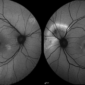
Widefield fundus autofluorescence of a 60-year-old male with Central Serous Chorioretinopathy affecting both eyes. Chronic history of CSR followed with observation without treatment prior to presenting at our office. The physician noted significant findings on exam and imaging with multifocal areas of inactive and active changes in the right eye and subfoveal subretinal fluid with recent visual decline in the left eye. There are hyper and hypoautofluorescent changes, consistent with CSR.
Photographer: Olivia Rainey, OCT-C, COA
Imaging device: Heidelberg Spectralis
Condition/keywords: 55-degrees, central serous chorioretinopathy (CSCR), central serous retinopathy (CSR), chronic central serous chorioretinopathy (CSCR), fundus autofluorescence (FAF), heidelberg spectralis, left eye
-
 Central Serous Chorioretinopathy
Jan 25 2022 by Olivia Rainey
Late phase widefield fluorescein angiography of a 60-year-old male with Central Serous Chorioretinopathy. Chronic history of CSR followed with observation without treatment prior to presenting at our office. The physician noted subfoveal subretinal fluid with recent visual decline. FA shows multifocal leakage and ICG shows hypercyanescence. OCTA, ICG, and FA consistent with CSR, and without concern for CNVM thus will observe without anti-VEGF at this time. PDT therapy recommended.
Photographer: Olivia Rainey, OCT-C, COA
Imaging device: Heidelberg Spectralis
Condition/keywords: 55-degrees, central serous chorioretinopathy (CSCR), central serous retinopathy (CSR), chronic central serous chorioretinopathy (CSCR), fluorescein angiogram (FA), heidelberg spectralis, indocyanine green (ICG) angiography, left eye
-
 Choroidal Detachment
Jan 17 2022 by Logan ryzenga
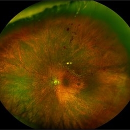
Left ultra-wide field photograph of an 81-year old female with a choroidal detachment affecting her left eye. Patient had a stent placed November, 2021 and following the procedure she complains of variable blurred vision and severe constricted visual fields. She presented at our office with flashes a month prior but without pain or floaters.
Photographer: Logan Ryzenga
Imaging device: Optos California
Condition/keywords: choroidal detachment, fundus photograph, left eye, Optos, pseudocolor, superior retina, ultra-wide field imaging
-
 Choroidal Detachment OS
Jul 5 2024 by Zach Seim
Optos Fundus Photograph of a Choroidal Detachment OS in a 75 year old male. VA at presentation was DCC HM.
Photographer: Zach Seim
Imaging device: Optos California
Condition/keywords: choroidal detachment, choroidal mass, left eye, optos, OPTOS CALIFORNIA
-
 Choroidal Hemangioma
Jan 19 2021 by Stacie Neview
Ultra wide field fluorescein angiogram of a 44-year-old male with presumed central serous retinopathy. Based on extended ophthalmoscopy, diagnostic ocular ultrasonography, and retinal imaging, the choroidal tumor is most consistent with a choroidal hemangioma. A circumscribed choroidal hemangioma such as this one is unlikely to be associated with an underlying systemic condition and will be further monitored and assessed for possible treatment.
Photographer: Stacie Neview, COA, OSC
Imaging device: Optos California
Condition/keywords: choroidal hemangioma, early phase, fluorescein angiogram (FA), left eye, Optos, ultra-wide field imaging
-
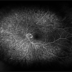
 Choroidal Melanoma with a Serous Retinal Detachment
Apr 30 2019 by Olivia Rainey
Ultra-wide field fundus autofluorescence of a 67-year-old male with serous retinal detachment, secondary to a large choroidal melanoma affecting the left eye. Patient reported a curtain affecting his superior field for about 2-3 months prior to examination. Patient elects radiation bracytherapy with a guarded visual expectation secondary to the location of the tumor, touching the optic disc.
Photographer: Olivia Rainey
Imaging device: Optos
Condition/keywords: autofluorescence imaging, left eye, Optos, ultra-wide field imaging
-
 Choroideremia
Sep 21 2022 by Zach Seim
Ultra-widefield fundus photo of a 74 year old male presenting with severe vision loss beginning at age 55. Patient sought a second opinion with our office and was diagnosed with Choroideremia. Patient denies hearing loss, heart problems, balance issues, polydactyly, kidney problems, and dental problems. Patient reports that nobody in the family had blindness. Choroideremia is an X-linked chorioretinal dystrophy characterized by the diffuse, progressive degeneration of the retinal pigment epithelium (RPE), photoreceptors and choriocapillaris. It is caused by a mutation in the CHM gene.
Photographer: Zach Seim
Imaging device: Optos California
Condition/keywords: choroideremia, hereditary choroidal atrophy, hereditary retinal dystrophy, left eye, light perception, low vision, Optos, pseudocolor, ultra-wide field imaging
 Loading…