 Initializing download.
-
 By Stacie Neview
Retina Specialists of Michigan - Uploaded on Jan 19, 2021.
- Last modified by Caroline Bozell on Jan 19, 2021.
- Rating
- Appears in
- Miscellaneous
- Condition/keywords
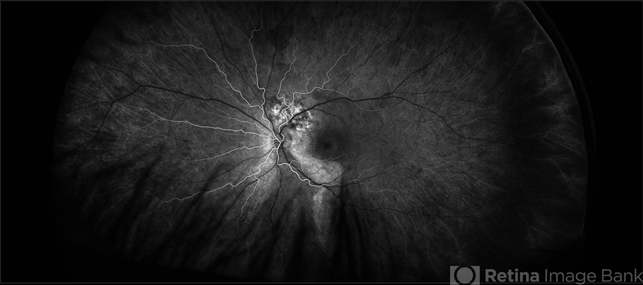
- choroidal hemangioma, ultra-wide field imaging, fluorescein angiogram (FA), Optos, left eye, early phase
- Photographer
- Stacie Neview, COA, OSC
- Imaging device
-
Fundus camera
Optos California - Description
- Ultra wide field fluorescein angiogram of a 44-year-old male with presumed central serous retinopathy. Based on extended ophthalmoscopy, diagnostic ocular ultrasonography, and retinal imaging, the choroidal tumor is most consistent with a choroidal hemangioma. A circumscribed choroidal hemangioma such as this one is unlikely to be associated with an underlying systemic condition and will be further monitored and assessed for possible treatment.


