Search results (119 results)
-
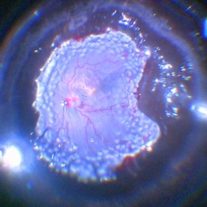 360 Degree Retinectomy
Feb 2 2022 by Manish Nagpal, MD, FRCS (UK), FASRS
Intraoperative photo of a case of retinal detachment with extensive PVR, which underwent 360 degree relaxing retinectomy followed by 360 laser barrage just prior to silicone oil injection.
Photographer: Manish Nagpal, Retina Foundation, Ahmedabad, India
Imaging device: Sony PMW -10 MD surgical camera
Condition/keywords: laser, laser photocoagulation, proliferative vitreoretinopathy (PVR), relaxing retinectomy, retinectomy, silicone oil
-
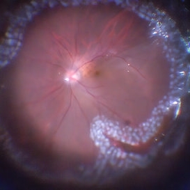 360 Endolaser Barrage
Feb 2 2022 by Manish Nagpal, MD, FRCS (UK), FASRS
Intraoperative photo of a 360 laser barrage done for a case of retinal detachment with a large superior tear.
Photographer: Manish Nagpal, Retina Foundation, Ahmedabad, India
Imaging device: Sony PMW -10 MD surgical camera
Condition/keywords: laser, laser photocoagulation, tear
-
 ARMD - CNV and Subretinal Fluid Post Thermal Laser
Aug 7 2013 by H. Michael Lambert, MD
ARMD - CNV post thermal laser.
Condition/keywords: choroidal neovascularization (CNV), laser photocoagulation
-
 Branch Retinal Vein Occlusion s/p Laser Treatment
Apr 8 2019 by Gary R. Cook, MD, FACS
65-year-old white male immediately status post quadrantic scatter and grid macular laser photocoagulation treatment of a nonperfused superotemporal BRVO with macular edema OD; V.A. = 20/50
Imaging device: Topcon VT-50
Condition/keywords: branch retinal vein occlusion (BRVO), laser photocoagulation, macular edema
-
 Choroidal Hemangioma
Mar 27 2019 by Gary R. Cook, MD, FACS
Immediate S/P argon laser photocoagulation of the choroidal hemangioma in a 36-year-old white male; V.A.= counting fingers at 4 feet.
Imaging device: Topcon VT-50
Condition/keywords: choroidal hemangioma, exudative detachment, laser photocoagulation, secondary retinal detachment
-
 Coats' Disease
Aug 24 2018 by Kim Barrett
Montage fluorescein angiography of 14-year-old male with Coats' Disease of the left eye. Multiple focal laser treatments. Current uncorrected visual acuity is 20/15-1 OU.
Photographer: Kim Barrett, C.O.A. Retina Specialist of Michigan
Imaging device: Heidelberg Spectralis
Condition/keywords: adolescent, Coats' disease, fluorescein angiogram (FA), Heidelburg Spectralis, laser photocoagulation, left eye, macroaneurysm, montage
-
 CSR Treated with Focal Laser: FFA
Dec 6 2021 by Nizamuddin HM Shaik, MD, FRCS
FFA of 35-year-old lady with CSR treated with focal laser.
Photographer: Mahmoud , Ophthalmology Technecian, International Medical Center
Imaging device: OCT
Condition/keywords: central serous chorioretinopathy (CSCR), FFA, focal laser, laser photocoagulation
-
 CSR Treated with Focal Laser: Fundus Photo
Dec 6 2021 by Nizamuddin HM Shaik, MD, FRCS
Fundus photograph of 35-year old lady with CSR treated with focal laser.
Photographer: Mahmoud , Ophthalmology Technician, International Medical Center
Imaging device: OCT
Condition/keywords: central serous chorioretinopathy (CSCR), focal laser, laser photocoagulation
-
 CSR Treated with Focal Laser: Fundus, FFA, OCT Images
Dec 6 2021 by Nizamuddin HM Shaik, MD, FRCS
Fundus photograph , FFA and OCT ( Pre and Post ) of a 35-year-old lady with CSR treated with focal laser.
Photographer: Mahmoud , Ophthalmology Technician, International Medical Center
Imaging device: OCT
Condition/keywords: central serous chorioretinopathy (CSCR), laser photocoagulation
-
 Eales Disease
Apr 1 2019 by Gary R. Cook, MD, FACS
23-year-old Vietnamese female status post peripheral laser panretinal photocoagulation (PRP) treatment showing regression of peripheral neovascularization with gliosis for Eales disease.
Imaging device: Topcon VT-50
Condition/keywords: Eales disease, gliosis, laser photocoagulation, pan-retinal photocoagulation (PRP)
-
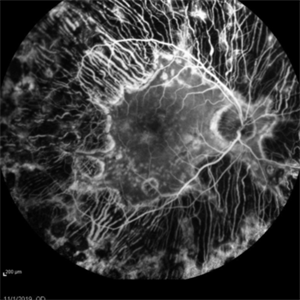
 Eals Disease
Jan 26 2013 by Ratimir Lazic, MD, PhD
FAG image of a 28-year-old male. Staining of scars due to laser photocoagulation can be seen.
Photographer: Marko Lukic, MD
Imaging device: Zeis Visucam Lite 2
Condition/keywords: fundus photograph, laser photocoagulation
-
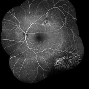
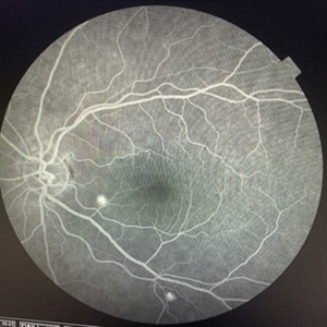
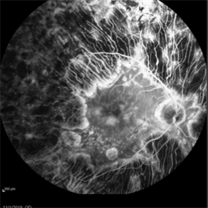 Enough PRP? (Proliferative Diabetic Retinopathy With Extreme PRP and Widespread Atrophy)
Nov 24 2019 by Thomas A. Ciulla, MD, MBA, FASRS
Fluorescein angiogram from a 71-year-old woman who underwent numerous sessions of pan retinal laser photocoagulation for proliferative diabetic retinopathy in the remote past. Note the widespread severe secondary atrophy, with only the central macular RPE remaining. Note the choroidal vessels through the diffuse window defect in the peripheral macula and near periphery.
Condition/keywords: laser injury, laser photocoagulation, proliferative diabetic retinopathy (PDR)
-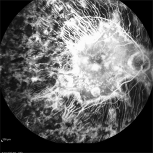
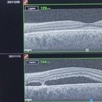
 Enough PRP? (Proliferative Diabetic Retinopathy With Extreme PRP and Widespread Atrophy)
Nov 24 2019 by Thomas A. Ciulla, MD, MBA, FASRS
OCT from a 71-year-old woman who underwent numerous sessions of pan retinal laser photocoagulation for proliferative diabetic retinopathy in the remote past. Note the widespread severe secondary atrophy, with only the central macular RPE remaining.
Condition/keywords: laser injury, laser photocoagulation, proliferative diabetic retinopathy (PDR)
-
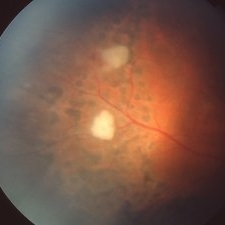
 Enough PRP? (Proliferative Diabetic Retinopathy With Extreme PRP and Widespread Secondary Atrophy)
Nov 24 2019 by Thomas A. Ciulla, MD, MBA, FASRS
Fundus photograph from a 71-year-old woman who underwent numerous sessions of pan retinal laser photocoagulation for proliferative diabetic retinopathy in the remote past. Note the widespread severe secondary atrophy, with only the central macular RPE remaining.
Condition/keywords: laser injury, laser photocoagulation, proliferative diabetic retinopathy (PDR)
-
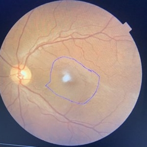
 Familial Exudative Vitreoretinopathy
Nov 25 2022 by Aditya S Kelkar, MS, FRCS, FASRS,FRCOphth
Colour fundus photograph of the right eye of a 56-year-old lady showing lasered FEVR with epiretinal membrane and vitreous band.
Photographer: Dr. Pranali Surawase. National Institute of Ophthalmology, Pune, Maharashtra, India
Imaging device: Zeiss Clarus 500
Condition/keywords: ERM, familial exudative vitreoretinopathy (FEVR), laser photocoagulation
-
 Fibrous Scaring After Laser Photocoagulation
Apr 2 2019 by Gary R. Cook, MD, FACS
Elderly white female with extensive fibrous scarring and worsened atrophy following focal Argon laser photocoagulation X 2 for exudative AMD; V.A. = 20/200
Imaging device: Topcon VT-50
Condition/keywords: complication, exudative age-related macular degeneration, fibrous macular scar, laser photocoagulation, laser surgery complications
-
 Floating Ozurdex Implant
May 20 2024 by Tejaswita Verma
Fundus photograph of the left eye of a 73 year old female with ozurdex implant floating in the vitreous in a diabetic lasered patient.
Photographer: DR. TEJASWITA VERMA
Imaging device: MIRANTE
Condition/keywords: diabetic macular edema, laser photocoagulation, Ozurdex implant
-
 Focal Laser Treatment for Central Serous Retinopathy: FFA
Dec 6 2021 by Nizamuddin HM Shaik, MD, FRCS
FFA of a 35-year-old lady with CSR treated with focal laser.
Photographer: Mahmoud , Ophthalmology Technecian, International Medical Center
Imaging device: OCT
Condition/keywords: central serous chorioretinopathy (CSCR), laser photocoagulation
-
 Fundus Photo Macular Choroidal Hemangioma Treated with Laser
Nov 11 2019 by Sophia El Hamichi, MD
A 51-year-old female that presented with a macular choroidal hemagioma complicated by focal exudative retinal detachment OD. The patient was treated with vitrectomy and laser therapy of the choroidal hemagioma along with bevacizumab intravitreal injection during and after the surgery. The patient evolved well with resolution of the subretinal fluid OD. VA 20/200
Photographer: Sophia El Hamichi,MD, Murray Ocular Oncology and Retina, Miami
Condition/keywords: laser photocoagulation, subretinal fluid
-
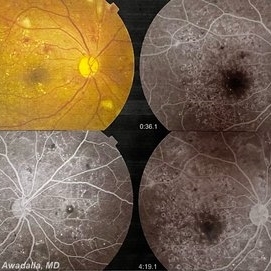 Grid macular photocoagulation for DME
Feb 17 2023 by Mohamed Awadalla
A case of aggressive grid macular photocoagulation treatment for DME. The treating ophthalmologist had done almost "pan-macular" photocoagulation
Condition/keywords: Grid macular photocoagulation, laser photocoagulation
-
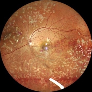
 High risk Proliferative Diabetic Retinopathy treated with Pan Retinal Photocoagulation
Nov 5 2022 by Somnath Chakraborty, MD
A Fundus Photo Montage of 43 year old Asian Male with Type 2 Diabetes Mellitus since 7 years who presented with sudden onset diminition of vision in his Left eye. BCVA OS was 20/200. He was diagnosed to have Pre retinal bleed due to Proliferative Diabetic Retinopathy and was treated with Pan Retinal Photocoagulation. This image shows a large neo-cascular frond at the disc and superior to it with Pre-retinal bleed and Fresh laser marks along
Photographer: Pulak Roy
Condition/keywords: diabetic blindness, diabetic retinopathy vitrectomy study (DRVS), fresh laser burns, laser photocoagulation, preretinal hemorrhage, proliferative diabetic retinopathy (PDR)
-
 Idiopathic Retinal Vasculitis
Jun 9 2024 by Anjana Mirajkar, MS Ophthalmology
A widefield image of a 32 year old male of LE showing laser marks in inferior and superior half with an floating ozurdex implant (inferiorly) in a case of idiopathic retinal vasculitis.
Photographer: Dr. Anjana Mirajkar -Retina Foundation, Ahmedabad
Imaging device: Mirante-Nidek
Condition/keywords: idiopathic retinal vasculitis, laser photocoagulation, Ozurdex implant, pan-retinal photocoagulation (PRP)
-
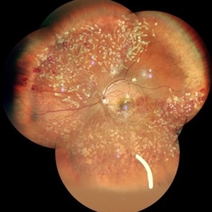 Idiopathic Retinal Vasculitis
Jun 9 2024 by Anjana Mirajkar, MS Ophthalmology
A color photo montage of an 32 year old male of LE showing laser marks in inferior and superior half with an floating ozurdex implant (inferiorly) in a case of idiopathic retinal vasculitis.
Photographer: Dr. Anjana Mirajkar -Retina Foundation, Ahmedabad
Imaging device: Mirante-Nidek
Condition/keywords: idiopathic retinal vasculitis, laser photocoagulation, Ozurdex implant
 Loading…