Search results (57 results)
-
 Angioid Streaks With Associated Disc Drusen and CNV
Sep 21 2018 by Sarah Oelrich
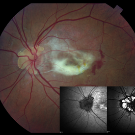
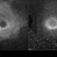
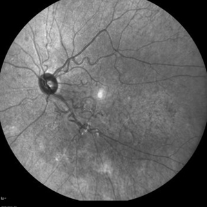
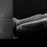
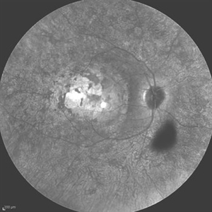
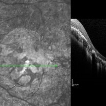
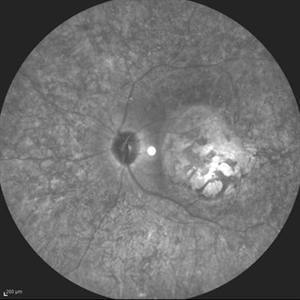
Angioid streaks with associated disc drusen and CNV.
Photographer: Sarah Oelrich CRA COT, Southeastern Retina Associates Knoxville Tn
Condition/keywords: angioid streaks, autofluorescence imaging, choroidal neovascularization (CNV), disc drusen, infrared image
-
 Behcet's Disease
Mar 13 2013 by Hamid Ahmadieh, MD
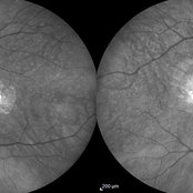
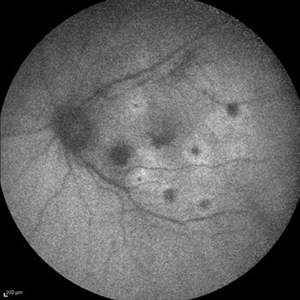
Infrared image of the right eye of a 23-year-old man with retinal vasculitis and branch retinal vein occlusion (BRVO) due to Behcet's disease .
Photographer: Solmaz Shahmohammad, Negah Eye Center, Tehran
Imaging device: Heidelberg Spectralis
Condition/keywords: branch retinal vein occlusion (BRVO), infrared image, retinal vasculitis
-
 Best Disease
Mar 9 2013 by Hamid Ahmadieh, MD
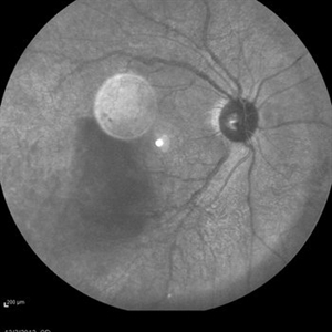
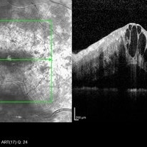
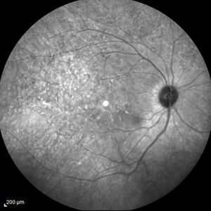
Infrared imaging of the left eye of a 49-year-old man with decreased VA due to advanced Best disease.
Photographer: Soodabeh Fooladin, Negah Eye Center, Tehran
Imaging device: Heidelberg Spectralis
Condition/keywords: Best disease, infrared image
-
 Case 2 Retinitis Pigmentosa BAF IRAF OD
May 14 2014 by Avris Romario Diparaja Siahaan
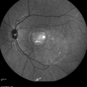
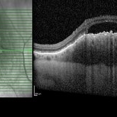
Fundus image a 57-year-old man with retinitis pigmentosa on both eyes. These image were taken with blue auto fluorescein mode (BAF) and infrared auto fluorescence (IRAF).
Photographer: Avris Romario Diparaja Siahaan
Imaging device: Heidelberg HRA + OCT Spectralis
Condition/keywords: autofluorescence imaging, fundus photograph, infrared image, retinitis pigmentosa
-
 Case 2 Retinitis Pigmentosa BAF IRAF OS
May 14 2014 by Avris Romario Diparaja Siahaan
Fundus image a 57-year-old man with retinitis pigmentosa on both eyes. These image were taken with blue auto fluorescein mode (BAF) and infrared auto fluorescence (IRAF).
Photographer: Avris Romario Diparaja Siahaan
Imaging device: Heidelberg HRA + OCT Spectralis
Condition/keywords: autofluorescence imaging, fundus photograph, infrared image, retinitis pigmentosa
-
 Central Areolar Choroidal Dystrophy
Jul 7 2015 by Hamid Ahmadieh, MD
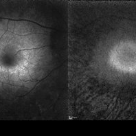
Infrared image of both eyes of a 58-year-old man with progressive loss of vision. VA OD is 20/60 and VA OS is 20/400.
Photographer: Soulmaz Shahmohammad, Negah Eye Center, Tehran, Iran
Imaging device: Specteralis
Condition/keywords: central areolar choroidal dystrophy (CACD), infrared image
-
 Central Retinal Vein Occlusion
Oct 7 2015 by Avris Romario Diparaja Siahaan
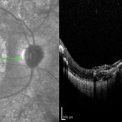
An IR + OCT image of a 46-year-old man with a central retinal vein occlusion on his left eye.
Photographer: Avris Romario Diparaja Siahaan, Klinik Mata Nusantara
Imaging device: Spectralis Heidelberg
Condition/keywords: central retinal vein occlusion (CRVO), infrared image, optical coherence tomography (OCT)
-
 Choroidal Melanoma
Feb 2 2018 by Olivia Rainey
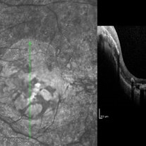
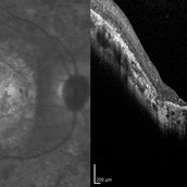
Optical coherence tomography with enhanced depth imaging of a 78-year-old female with choroidal melanoma with subretinal fluid affecting her right eye.
Photographer: Olivia Rainey
Imaging device: Heidelberg Spectralis
Condition/keywords: enhanced depth imaging, infrared image, optical coherence tomography (OCT), subretinal fluid, superior retina
-
 Combined Hamartoma of the Retina and Retinal Pigment Epithelium (CHRRPE)
Jan 21 2020 by Pierre-Henry Gabrielle, MD
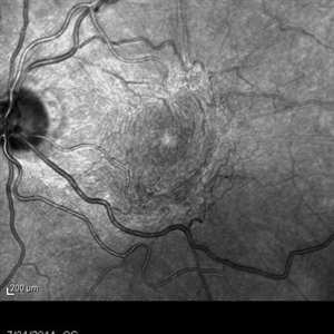
IR imaging of a 17-year-old man with Combined hamartomas of the retina and retinal pigment epithelium (CHRRPE) at the posterior pole of the left eye.
Photographer: Pierre-Henry Gabrielle, Ophthalmology department, Dijon University Hospital, France
Imaging device: Heidelberg Spectralis
Condition/keywords: combined hamartoma, infrared image
-
 Cone-Rod Dystrophy
Mar 15 2017 by Hamid Ahmadieh, MD
Infrared and OCT images of the left eye of a 16-year-old boy with decreased visual acuity and color vision deficiency due to cone-rod dystrophy.
Photographer: Abazarnezhad , Negah Eye Center, Tehran, Iran
Imaging device: Spectralis OCT
Condition/keywords: cone dystrophy, infrared image, optical coherence tomography (OCT)
-
 Cone-Rod Dystrophy
Mar 15 2017 by Hamid Ahmadieh, MD
Infrared and OCT images of the right eye of a 16-year-old boy with decreased visual acuity and color vision deficiency due to cone-rod dystrophy.
Photographer: Abazarnezhad , Negah Eye Center, Tehran, Iran
Imaging device: Spectralis OCT
Condition/keywords: cone dystrophy, infrared image, optical coherence tomography (OCT)
-
CSNB-OCT-OD
Aug 17 2021 by Christine Kay, MD
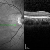
This is an OCT/infrared image OD exhibiting normal fundus in a 16 year-old male with X-linked CSNB with proven mutation in CACNA1F.
Photographer: Christine Kay, MD
Condition/keywords: infrared image, X-linked CSNB
-
 CSNB-OCT-OD
Aug 23 2021 by Jennifer Carstens
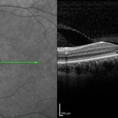
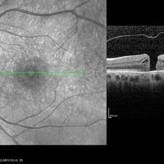
OCT/infrared image showing myopic fundus with normal retinal structure in patient with CACNA1F associated X-linked CSNB (OD).
Photographer: Jing Zhang, Ophthalmic Photographer
Condition/keywords: congenital stationary night blindness (CSNB), infrared image, optical coherence tomography (OCT)
-
 CSNB-OCT-OS
Aug 23 2021 by Jennifer Carstens
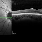
OCT/infrared image showing myopic fundus with normal retinal structure in patient with CACNA1F associated X-linked CSNB (OS).
Photographer: Jing Zhang, Ophthalmic Photographer
Condition/keywords: congenital stationary night blindness (CSNB), infrared image, optical coherence tomography (OCT)
-
 Endogenous Endophthalmitis
Sep 3 2014 by Hamid Ahmadieh, MD
Infrared image of the left eye of a 45-year-old diabetic man with the history of urinary tract infection. The most probable diagnosis was candida endogenous endophthalmitis.
Photographer: Nayereh Hadipour, Negah Eye Center, Tehran, Iran
Condition/keywords: candida endophthalmitis, endogenous endophthalmitis, infrared image
-
 Fundus Flavimaculatus and CNV
Nov 14 2013 by Hamid Ahmadieh, MD
Infrared image of the right eye of a 35-year-old woman with subfoveal CNV secondary to fundus flavimaculatus .
Photographer: Nayereh Hadipour, Negah Eye Center, Tehran
Condition/keywords: choroidal neovascularization (CNV), fundus flavimaculatus, infrared image, retinal flecks
-
 Infrared Macular Pucker
Dec 5 2014 by Stuart Alfred, CRA, OCT-C
Infrared macular pucker.
Photographer: Stuart Alfred, CRA, OCT-C
Condition/keywords: infrared image, macular pucker
-
 Iris Vascular Tuft
Jul 5 2022 by Olivia Rainey
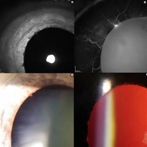
Anterior segment imaging of a 66-year-old male with Vascular Disorders of Iris and Ciliary Body affecting his right eye. The physician stated that the findings are most consistent with a benign vascular tuft at the pupillary margin. The patient presented at the office with 20/20 vision in both eyes and had no ocular complaints at the time of his appointment.
Photographer: Olivia Rainey, OCT-C, COA
Imaging device: Heidelberg Spectralis, Slit Lamp with Samsung Galaxy 7
Condition/keywords: anterior segment, fluorescein angiogram (FA), heidelberg spectralis, infrared image, near infrared autofluorescence (NIRAF), slit lamp photo, vascular anomaly, vascular disorders of iris and ciliary body, vascular tuft
-
 Leber's Congenital Amaurosis
Feb 25 2017 by Hamid Ahmadieh, MD
Infrared image of the right eye of a 25-year-old woman with bilateral macular colobomata and pigmentary retinopathy similar to Leber's congenital amaurosis.
Photographer: Shabnam Poureh, Negah Eye Center, Tehran, Iran
Condition/keywords: bilateral pigmentary retinopathy, infrared image, macular coloboma
-
 Macular Coloboma and Pigmentary Retinopathy
Feb 25 2017 by Hamid Ahmadieh, MD
Infrared and OCT images of the right eye of a 25-year-old woman with bilateral macular colobomata and pigmentary retinopathy similar to Leber's congenital amaurosis.
Photographer: Shabnam Poureh, Negah Eye Center, Tehran, Iran
Condition/keywords: infrared image, macular coloboma, optical coherence tomography (OCT)
-
 Macular Coloboma and Pigmentary Retinopathy
Feb 25 2017 by Hamid Ahmadieh, MD
Infrared and OCT images of the left eye of a 25-year-old woman with bilateral macular colobomata and pigmentary retinopathy similar to Leber's congenital amaurosis.
Photographer: Shabnam Poureh, Negah Eye Center, Tehran, Iran
Imaging device: Spectralis OCTc
Condition/keywords: infrared image
-
 Macular Coloboma and Pigmentary Retinopathy
Feb 25 2017 by Hamid Ahmadieh, MD
Infrared and OCT images of the left eye of a 25-year-old woman with bilateral macular colobomata and pigmentary retinopathy similar to Leber's congenital amaurosis.
Photographer: Shabnam Poureh, Negah Eye Center, Tehran, Iran
Condition/keywords: bilateral pigmentary retinopathy, infrared image, macular coloboma, optical coherence tomography (OCT)
-
 Macular Coloboma and Pigmentary Retinopathy
Feb 25 2017 by Hamid Ahmadieh, MD
Infrared image of the left eye of a 25-year-old woman with bilateral macular colobomata and pigmentary retinopathy similar to Leber's congenital amaurosis.
Photographer: Shabnam Poureh, Negah Eye Center, Tehran, Iran
Condition/keywords: bilateral pigmentary retinopathy, infrared image, macular coloboma
-
 Macular Coloboma and Pigmentary Retinopathy
Feb 25 2017 by Hamid Ahmadieh, MD
Infrared and OCT images of the right eye of a 25-year-old woman with bilateral macular colobomata and pigmentary retinopathy similar to Leber's congenital amaurosis.
Photographer: Shabnam Poureh, Negah Eye Center, Tehran, Iran
Imaging device: Spectralis OCT
Condition/keywords: infrared image, macular coloboma, optical coherence tomography (OCT)
-
 Macular Hole
Jul 26 2014 by Avris Romario Diparaja Siahaan
An OCT image of a 55-year-old-woman with macular hole on her right eye.
Photographer: Avris Romario Diparaja Siahaan, Klinik Mata Nusantara
Imaging device: Heidelberg Spectralis
Condition/keywords: infrared image, macular hole, optical coherence tomography (OCT)
 Loading…