Search results (15 results)
-
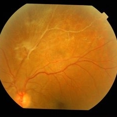
 Eales Disease
Apr 1 2019 by Gary R. Cook, MD, FACS
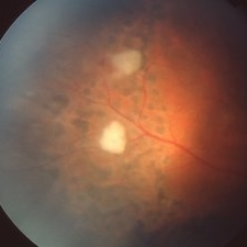
23-year-old Vietnamese female status post peripheral laser panretinal photocoagulation (PRP) treatment showing regression of peripheral neovascularization with gliosis for Eales disease.
Imaging device: Topcon VT-50
Condition/keywords: Eales disease, gliosis, laser photocoagulation, pan-retinal photocoagulation (PRP)
-
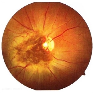
 Slide 13-13
Mar 4 2019 by Lancaster Course in Ophthalmology
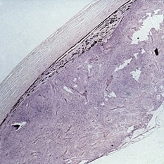
Retinoblastoma and gliosis of the retina overlying the optic nerve ( x25).
Condition/keywords: gliosis, retinoblastoma
-
 Slide 7-81
Feb 25 2019 by Lancaster Course in Ophthalmology
Massive retinal gliosis.
Condition/keywords: gliosis
-
 Slide 9-30
Feb 26 2019 by Lancaster Course in Ophthalmology
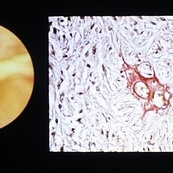
Massive gliosis of the retina (clinicopathologic correlation). Lesion consists of uniform spindle-shaped cells, abundant fibrillar material, and sclerotic blood vessels.
Condition/keywords: clinicopathologic correlation, gliosis
-
 Slide 9-89
Feb 26 2019 by Lancaster Course in Ophthalmology
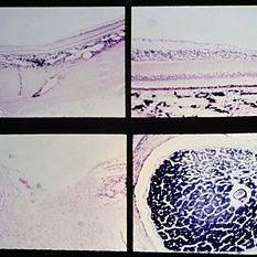
Nutritional amblyopia. The nerve fiber and ganglion cell layers are absent in the macular area (upper views). The temporal side of the optic nerve head (lower left) is partially atrophy, with marked reduction in the size of the nerve fiber bundles and secondary gliosis.
Condition/keywords: amblyopia, atrophy, gliosis, macular
-
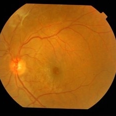
 Branch Retinal Vein Occlusion With Proliferative Retinopathy
Jul 29 2014 by Mallika Goyal, MD
Left eye of a 54-year-old visually asymptomatic male shows superonasal BRVO with proliferation and gliosis on routine fundus exam.
Photographer: Mallika Goyal, MD, Apollo Health City, Jubilee Hills, Hyderabad-500033
Condition/keywords: branch retinal vein occlusion (BRVO)
-
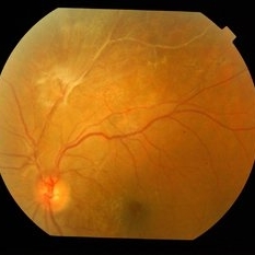 BRVO With Proliferative Retinopathy
Sep 22 2014 by Mallika Goyal, MD
Left fundus of 54-year-old male shows ST BRVO with preretinal gliosis suggestive of proliferative retinopathy.
Photographer: Mallika Goyal, MD, Apollo Health City, Jubilee Hills, Hyderabad-500033
Condition/keywords: branch retinal vein occlusion (BRVO)
-
 Capillary Hemangioma
Mar 27 2019 by Gary R. Cook, MD, FACS
Left eye of a 35-year-old white female with a capillary hemangioma of the optic disc OS; V.A.= 20/40. This hemangioma showed significant preretinal gliosis, and was not associated with von Hippel-Lindau disease.
Condition/keywords: hemangioma
-
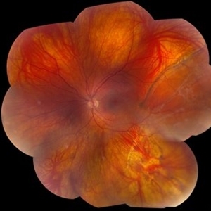 Chronic Retinal Detachment in a Young Myopic Patient
Nov 6 2019 by Kamal Kishore, MD, MBBS
Chronic retinal detachment in a 27-year-old myopic female showing spontaneous reattachment in inferotemporal quadrant, and demarcation line and subretinal gliosis in superotemporal quadrant.
Photographer: Stephanie Shaver
Imaging device: Topcon 50 EX with OIS Winstation
Condition/keywords: chronic retinal detachment, high myopia
-
 Combined Hamartoma of the Retina and Retinal Pigment Epithelium
Apr 26 2020 by Dipak Nag, MBBS, FCPS, MSc, FRF
A 28-year-old female presented with a deeply pigmented gray- brown, elevated lesion extending from the temporal side of the disc to the macula (OU). Remarkable retinal vasculature with straightening of the distal vessels and dilatation as well as tortuousity of the peri-lesional vessels. The vitreoretinal interface shows gliosis and epi-retinal membrane (ERM) formation.
Photographer: Dipak Nag
Condition/keywords: hamartoma, retinal pigment epithelium
-
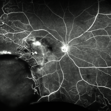 Fluorescein Angiography (FA) of a Primary Retinal Vasoproliferative Tumor
Jun 29 2025 by Marcelo Zas, MD PhD
We present a case of a 33-year-old male patient, who presented with decreased visual acuity in his right eye with 20/80, presenting a primary retinal vasoproliferative tumor in the lower temporal quadrant. The fluorescein angiography findings are: 1. Early hyperfluorescence due to its rich intrinsic vascularity and often has dilated feeding arterioles and draining venules. 2. Marked progressive leakage from the tumor vessels. 3. The late leakage often obscures fine vascular details in the late phase and corresponds to exudation and macular edema seen clinically. 4. Staining of surrounding exudates, RPE disturbances and gliosis. 5. In our case also a marked peripheral capillary closure in the areas adjacent to the tumor and in other quadrants as well.
Photographer: Marcelo Zas MD PhD
Condition/keywords: RETINAL VASOPROLIFERATIVE TUMOR
-
 Spontaneously Attached Retina
Jan 15 2021 by KRISHNENDU NANDI, MS
Fundus photograph of 26-year-old man with BCVA 6/60, N24, showed spontaneous retinal reattachment with multiple retinal cyst at temporal quadrant. Subretinal gliosis resemble a mustache.
Photographer: Krishnendu Nandi, Netralayam Eye Care Centre
Condition/keywords: spontaneous retinal reattachment
 Loading…