Search results (55 results)
-
 Gas Bubble
May 21 2021 by Raj K. Maturi, MD
S/P PPV 73-year-old woman with small retinal detachment. Originally presented with decrease vision and residual vitreous floaters. Upon examination small localized retinal detachment with multiple breaks.
Photographer: Charlotte Harris ,Midwest Eye Institute 10300 N Illinois St, Carmel Indiana 46290
Imaging device: Nikon Optos California P200TDx
Condition/keywords: gas bubble
-
 Gas Bubble
May 21 2021 by Raj K. Maturi, MD
S/P PPV 73-year-old woman with small retinal detachment. Originally presented with decrease vision and residual vitreous floaters. Upon examination small localized retinal detachment with multiple breaks.
Photographer: Charlotte Harris ,Midwest Eye Institute 10300 N Illinois St, Carmel Indiana 46290
Imaging device: Nikon Optos California P200TDx
Condition/keywords: gas bubble
-
 Gas Bubble
May 21 2021 by Raj K. Maturi, MD
S/P PPV 73-year-old woman with small retinal detachment. Originally presented with decrease vision and residual vitreous floaters. Upon examination small localized retinal detachment with multiple breaks.
Photographer: Charlotte Harris ,Midwest Eye Institute 10300 N Illinois St, Carmel Indiana 46290
Imaging device: Nikon Optos California P200TDx
-
---thumb.JPG/image-square;max$300,300.ImageHandler) Gas Bubble
Gas Bubble
Jul 12 2013 by Jason S. Calhoun
Patient had a pneumatic retinopexy for retinal detachment. Fundus shows C3F8 gas bubble superiorly.
Photographer: Jason S. Calhoun, Department of Ophthalmology, Mayo Clinic Jacksonville, Florida
Condition/keywords: pneumatic retinopexy
-
 Gas Bubble Extending into Anterior Chamber
Oct 12 2012 by Jeffrey G. Gross, MD, FASRS
Gas bubble extending into anterior chamber in aphakic eye, after PPV.
Condition/keywords: anterior chamber, aphakic eye, gas bubble, pars plana vitrectomy (PPV)
-
 Gas Bubble in Anterior Chamber
Jul 14 2013 by Jason S. Calhoun
Gas bubble injected in anterior chamber to control IOP.
Photographer: Jason S. Calhoun, Department of Ophthalmology, Mayo Clinic Jacksonville, Florida
Imaging device: TOPCON D-90 SL NIKON CAMERA
Condition/keywords: gas bubble
-
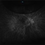
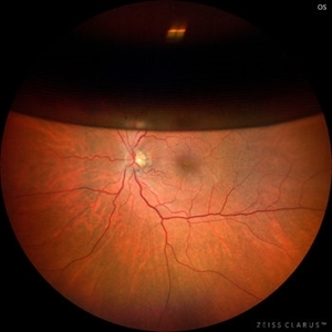
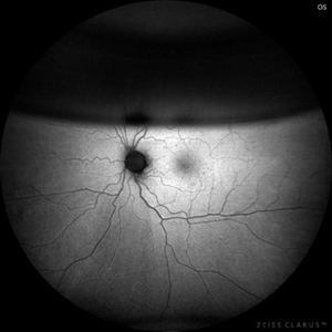 Intraocular Gas bubble - Wide field Fundus Autofluorescence
Sep 6 2021 by Ricardo Leitão Guerra
Wide field confocal scanning laser ophthalmoscopy 7 days after vitrectomy to treat a macular hole.
Imaging device: Zeiss Clarus 700
Condition/keywords: gas bubble, macular hole, pars plana vitrectomy (PPV), post-vitrectomy, vitrectomy
-
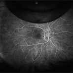
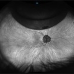
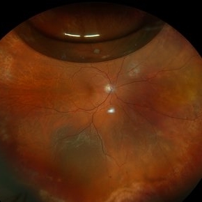
 Intraocular Gas Bubble - Wide-field True Color CSLO
Sep 6 2021 by Ricardo Leitão Guerra
Wide field confocal scanning laser ophthalmoscopy 7 days after vitrectomy to treat a macular hole.
Imaging device: Zeiss Clarus 700
Condition/keywords: gas bubble, macular hole, pars plana vitrectomy (PPV), post-vitrectomy, vitrectomy
-
 Macular Hole Stage 3
Sep 27 2012 by Jeffrey G. Gross, MD, FASRS
Macular hole stage 3 post op with gas bubble 20/60.
Condition/keywords: 20/60, gas bubble, macular hole, post-op
-
 Macular Hole With Trapped Gas Bubble
Jan 5 2018 by Manish Nagpal, MD, FRCS (UK), FASRS
Fundus photo of a patient who was operated for macular hole surgery had come for a follow up after one month and revealed a persisting macular hole with a small trapped gas bubble within the hole.
Photographer: rakesh juneja
Condition/keywords: gas bubble, macular hole
-
 New Retinal Detachment 6w s/p RD repair
Nov 16 2023 by Virginia Gebhart
13 year old male presented with new blind spot 6 weeks s/p RD repair with cryo/scleral buckle/prophylaxis laser with gas bubble. New RD involving the macula, posterior to scleral buckle, secondary to PVD. Small gas bubble remaining. Pt was brought back to OR for repeat PPV and silicone oil repair
Photographer: Virginia Gebhart
Imaging device: Optos
Condition/keywords: gas bubble, Retinal Detachment, retinal detachment of the macula, scleral buckle
-
 Pseudophakic RRD, S/P Buckle/Vit. w/ Residual Gas Fish Eggs OD
May 23 2018 by Hosam Attia, MD
71-year-old male, s/p combined buckle vitrectomy for recurrent, macula-off, rhegmatogenous retinal detachment, with residual gas fish eggs OD.
Imaging device: Optos California Ultra-Wide Field Fundus Camera
Condition/keywords: encircling scleral buckle, gas bubble, intraocular gas, intravitreal gas bubble
-
 Recurrent Retinal Detachment with Single Break
Nov 2 2024 by Virginia Gebhart
84 year old male with recurrent detachment s/p PPV/RD repair 2 weeks ago. Retinotomy is opened and appears to be the source of the fluid. Pt scheduled for emergency repair with scleral buckle.
Photographer: Virginia Gebhart
Imaging device: Optos California
Condition/keywords: gas bubble, retinal detachment, retinotomy
-
 Repaired Retinal Detachment with Grade C PVR
Dec 23 2024 by Virginia Gebhart
61 year old male 1 day s/p retinectomy/SO exchange. Retina is attached under SO with good laser to retinectomy edge.
Photographer: Virginia Gebhart, Retina Consultants of Carolina
Imaging device: Optos California
Condition/keywords: gas bubble, proliferative vitreoretinopathy (PVR), retinectomy, silicone oil, total retinal detachment
-
 Retinal Detachment with PVR
Feb 24 2025 by Kimberly Wakester
Optomap RGB of an 48-year-old man with a retinal detachment with PVR. Patient is 6 weeks s/p RD repair with giant HSRT. Patient has new PVR noted on post op exam causing the retina to re-detach. Patient is having to have a 2nd surgery to remove the scar tissue and have silicone oil placement. Will continue close follow up care.
Photographer: Kimberly Wakester, COA
Imaging device: Optos California
Condition/keywords: gas bubble, PVR, retinal detachment
-
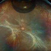
 T-Cell Lymphoma
Jul 3 2025 by Virginia Gebhart
78 year old male s/p vitreous biopsy for T-Cell lymphoma. Pt presented with peripheral blot hemorrhages and numerous white subretinal infiltrates. Retinal pallor and thickening temporally. History of cutaneous T-cell lymphoma. PPV/vitreous biopsy performed to find differential diagnosis. Silicone oil was placed for 6 weeks, then removed and exchanged with a gas bubble. Hematology pathologist and Emory reviewed path report and agrees it is consistent with T-cell lymphoma. Pt received intravitreal Methotrexate and will be scheduled for weekly treatments. BCVA CF
Photographer: Virginia Gebhart, Retina Consultants of Carolina
Imaging device: Optos California
Condition/keywords: biopsy, gas bubble, lymphoma
-
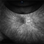
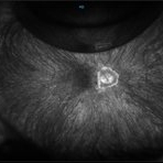
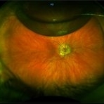
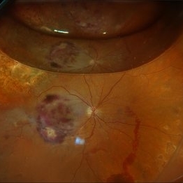
 Unexpected Sanctuary: Gas Bubble Entrapment in Morning Glory Disc
Sep 5 2025 by Danny Salgado Gómez
Fundus photograph of a 62-year-old male patient with Morning Glory syndrome in the right eye, who underwent vitrectomy, gas, and endolaser for posterior pole detachment. In the postoperative period, a gas bubble is observed within the optic disc, which persisted even after complete reabsorption of the intraocular gas.
Photographer: Dr. Danny Salgado, Retina and Vitreous Fellow, Clínica Oftalmológica del Caribe, Colombia.
Condition/keywords: gas bubble, intraocular gas, Morning Glory, Retinal Detachment, vitrectomy
-
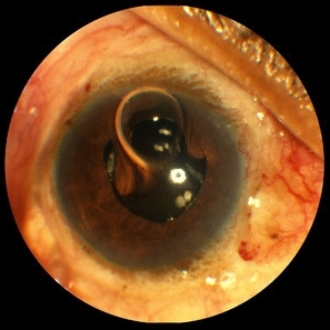
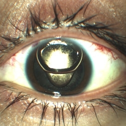 Anterior Chamber Gas and PFC Migration
Jun 21 2018 by Maria Stephanie R. Jardeleza, MD
Anterior segment photographs of 30-year-old male who underwent superior rhegmatogenous retinal detachment repair with intraocular gas tamponade. Perfluorocarbon was used to flatten the macula to prevent a macular fold and was removed during PFC/air exchange. Post operative week two visit shows gas migration into the anterior chamber with retained PFC layered in a tear drop shape posterior to the gas bubble and anterior to the lens. Patient had been maintaining face down positioning.
Photographer: Andy Zepeda, COA, Retina Clinic, San Antonio Eye Center, San Antonio, TX
Condition/keywords: retained perfluorocarbon, retina surgery complications, vitreous substitutes
-
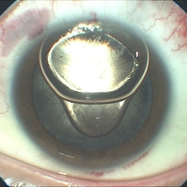 Anterior Segment Gas Bubble and PFC Interface
Jun 21 2018 by Maria Stephanie R. Jardeleza, MD
Anterior segment photographs of 30-year-old male who underwent superior rhegmatogenous retinal detachment repair with intraocular gas tamponade. Perfluorocarbon was used to flatten the macula to prevent a macular fold and was removed during PFC/air exchange. Post operative week two visit shows gas migration into the anterior chamber with retained PFC on the posterior aspect of the gas bubble/anterior surface of the lens. Patient had been maintaining face down positioning.
Photographer: Andy Zepeda, COA, Retina Clinic, San Antonio Eye Center, San Antonio, TX
Condition/keywords: retained perfluorocarbon, vitreous substitutes
 Loading…