Search results (2832 results)
-
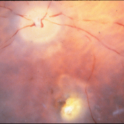
 Anterior segment
Feb 19 2013 by From the Collections of Thomas M. Aaberg, MD and Thomas M. Aaberg Jr., MD
Anterior segment photo taken with fundus camera.
Condition/keywords: fundus photograph, unknown
-
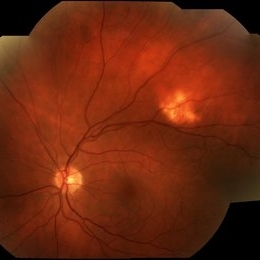
 ARMD With Geographic Atrophy, Peripheral Degeneration
Dec 6 2013 by James B. Soque, CRA, OCT-C, COA, FOPS
92-year-old white female with exudative macular degeneration, geographic atrophy, and peripheral retinal degeneration.
Photographer: James Soque, CRA COA, Island Retina, Shirley, New York
Imaging device: Topcon TRC 50DX with OIS 10.6.45
Condition/keywords: fundus photograph, geographic atrophy
-
 Best Vitelliform Macular Dystrophy
Dec 10 2020 by McGill University Health Centre
Postmortem eyes from 101-year-old female. Past clinical history includes a poor vision for many years due to macular degeneration. The last visual acuity test recorded 6/15 OD and 6/6 OS. IOP 14 and 18 torr OS. Histopathology: Disclosed and yellow 2x2mm macular lesion. Microscopic examination: elevated placoid macular lesion with overlying pigment granules. Electron microscopy examination: pigment granules with abundant lipofuscin and melanolysosomes, photoreceptor cells markedly attenuated (less degenerated at the periphery) Numerous calcified drusen throughout the retina particularly in the posterior pole. RPE lipofuscin content is elevated in Best’s dystrophy. The extractability of the PRE lipofuscin fluorophores is reduced (it is normal during senescence). The defect in Best’s dystrophy accelerates this age related change in lipofuscin.
Condition/keywords: Best vitelliform macular dystrophy (BVMD), fundus photograph
-
 Birdshot Retinopathy 5
Jan 15 2016 by Raj K. Maturi, MD
Fundus image of an 64-year-old white male.
Photographer: Tom Steele, CRA Midwest Eye Institute Indianapolis, Indiana
Imaging device: Optos
Condition/keywords: birdshot, fundus photograph
-
 Blunt Ocular Trauma Due to Firework Injury
Jun 9 2020 by Brittany Rota
Ultra- widefield pseudocolor image of an 18-year-old male with blunt ocular trauma in the right eye due to a firework injury. The patient presented with commotio retinae (sclopteria), an acute vitreous hemorrhage, choroidal rupture, and a subretinal hemorrhage. The referring physician performed surgery on the lateral rectus muscle which was macerated but not severed, and several orbital fibrous foreign bodies were removed from the posterior orbit. The globe was intact. There is no evidence of retinal tear in the region of sclopetaria; however, there is complete necrosis of the temporal peripheral choroid and retina. The vitreous hemorrhage was slowly clearing on his exam 6-9-2020. The patient is developing subretinal fibrosis. The physician is concerned about the choroidal rupture that is visible through the submacular hemorrhage. There is one rupture that appears to course directly under the fovea. The physician states that if this is the case, his vision most likely will be 20/200 or worse. His vision was hand motion in all fields except nasally, which he was unable to see hand motion at his visit on 6-9-2020.
Photographer: Brittany Rota
Imaging device: Optos California
Condition/keywords: blunt trauma, choroidal rupture, commotio retinae, fibrosis, firework injury, fundus photograph, hand motion, necrotizing retina, Optos, pseudocolor, subretinal hemorrhage, vitreous hemorrhage
-
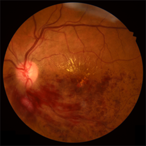
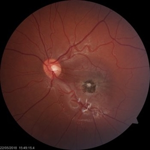
 Branch Retinal Artery Occlusion With Calcium Embolus at the Disc - Fundus Photo
Apr 7 2018 by Rameez N Hussain, MD
Acute branch retinal artery occlusion with a calcium embolus at the disc with retinal whitening in the area of retinal edema.
Photographer: DR RAMEEZ N HUSSAIN
Imaging device: zeiss
Condition/keywords: branch retinal artery occlusion (BRAO), embolus, fundus photograph, retinal edema
-
 Branch Retinal Artery Occlusion With Calcium Embolus at the Disc - Fundus Photo
Apr 7 2018 by Rameez N Hussain, MD
Acute retinal artery occlusion with a calcium embolus at the disc and retinal whitening.
Photographer: DR RAMEEZ N HUSSAIN
Imaging device: zeiss
Condition/keywords: branch retinal artery occlusion (BRAO), embolus, fundus photograph, retinal edema
-
 Branch Retinal Vein Occlusion
Apr 15 2023 by Yousef A Fouad, MD, FRCS (Glasg.)
BRVO in a young female with uncontrolled hypertension
Photographer: Yousef Fouad, Ain Shams University, Egypt
Condition/keywords: branch retinal vein occlusion (BRVO), fundus photograph, Hypertension, venous occlusion
-
 Case 2 Retinitis Pigmentosa BAF IRAF OD
May 14 2014 by Avris Romario Diparaja Siahaan
Fundus image a 57-year-old man with retinitis pigmentosa on both eyes. These image were taken with blue auto fluorescein mode (BAF) and infrared auto fluorescence (IRAF).
Photographer: Avris Romario Diparaja Siahaan
Imaging device: Heidelberg HRA + OCT Spectralis
Condition/keywords: autofluorescence imaging, fundus photograph, infrared image, retinitis pigmentosa
-
 Case 2 Retinitis Pigmentosa BAF IRAF OS
May 14 2014 by Avris Romario Diparaja Siahaan
Fundus image a 57-year-old man with retinitis pigmentosa on both eyes. These image were taken with blue auto fluorescein mode (BAF) and infrared auto fluorescence (IRAF).
Photographer: Avris Romario Diparaja Siahaan
Imaging device: Heidelberg HRA + OCT Spectralis
Condition/keywords: autofluorescence imaging, fundus photograph, infrared image, retinitis pigmentosa
-
 Cavernous Hemangioma of the Retina
Sep 20 2013 by Hector E. Ibanez, MD, FACS, FASRS, FABO, FAAO
Fundus photograph of an 8-year-old boy with a cavernous hemangioma of the retina.
Photographer: Hector E. Ibanez, MD, FACS
Imaging device: Topcon TRC 50
Condition/keywords: cavernous hemangioma of the retina, fundus photograph
-
 Central retinal vein obstruction (CRVO)
May 29 2022 by Eduardo Javier Pinuer Alvarado
Fundus photograph of a 63 year-old woman with central retinal vein obstruction (CRVO). The patient didn´t notice the decrease of VA, we found this in a routine exam for diabetic patients.
Photographer: Eduardo Pinuer A, Universidad Austral de Chile.
Imaging device: CR-2 AF Digital Non-Mydriatic Retinal Camera, Canon.
Condition/keywords: central retinal vascular obstruction, diabetes, fundus photograph
-
 Chorioretinal Scar
May 16 2017 by Olivia Rainey
Fundus photograph of an 17-year-old male with a macular scar affecting his right eye secondary to exudation from Coats disease.
Photographer: Olivia Rainey
Imaging device: Topcon 50dx
Condition/keywords: 20 degrees, chorioretinal scar, Coats' disease, color fundus photograph, color photo, fundus photograph
-
 Chorioretinitis
May 17 2014 by Avris Romario Diparaja Siahaan
Fundus photograph of a 34-year-old man with chorioretinitis (toxo) on his right eye.
Photographer: Prayid Listiyanto, Klinik Mata Nusantara
Imaging device: Topcon TRC 50 DX Type IA
Condition/keywords: fundus photograph, toxo chorioretinitis
-
 Choroidal Detachment
Jan 17 2022 by Logan ryzenga
Left ultra-wide field photograph of an 81-year old female with a choroidal detachment affecting her left eye. Patient had a stent placed November, 2021 and following the procedure she complains of variable blurred vision and severe constricted visual fields. She presented at our office with flashes a month prior but without pain or floaters.
Photographer: Logan Ryzenga
Imaging device: Optos California
Condition/keywords: choroidal detachment, fundus photograph, left eye, Optos, pseudocolor, superior retina, ultra-wide field imaging
-
 Choroidal Osteoma
Oct 11 2019 by Gayathri Mohan
Color fundus image of a peripapillary choroidal osteoma.
Photographer: Gayathri Mohan,Retina Foundation
Condition/keywords: choroidal osteoma, fundus photograph
-
---thumb.jpg/image-square;max$300,300.ImageHandler) Choroidal Osteoma
Choroidal Osteoma
Apr 29 2013 by Subijay Sinha
Fundus photograph of a 14 year old girl showing peripaillary amelanotic lesion with well defined margins.
Photographer: Subijay Sinha
Condition/keywords: fundus photograph
-
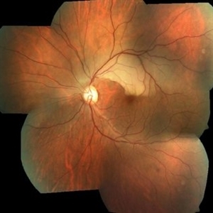
 Choroidal Osteoma Fundus Photograph Montage
Apr 17 2014 by Susanna S. Park, MD, PhD
Fundus photograph montage of a 72-year-old woman showing a mid-peripheral amelanotic lesion with minimal elevation which showed marked shadowing on ultrasonography consistent with a choroidal osteoma.
Photographer: Ellen Redenbo, University of California Davis Eye Center
Condition/keywords: choroidal osteoma, fundus photograph
-
 Choroideremia
Jan 26 2013 by Ratimir Lazic, MD, PhD
FAG image of a 66-year-old male. Diffuse chorioretinal atrophy is present. Large choroidal vessels can be seen. "Hyperflorescent" areas represent normal chorioretinal tissue.
Photographer: Marko Lukic, MD
Imaging device: Zeis Visucam Lite 2
Condition/keywords: choroideremia, fundus photograph
-
 Choroideremia
Jan 26 2013 by Ratimir Lazic, MD, PhD
FAG image of a 66-year-old male. Diffuse chorioretinal atrophy is present. Large choroidal vessels can be seen due to atrophy of the RPE and choriocapilaris.
Photographer: Marko Lukic, MD
Imaging device: Zeis Visucam Lite 2
Condition/keywords: choroideremia, fundus photograph
-
---thumb.jpg/image-square;max$300,300.ImageHandler) Choroideremia
Choroideremia
Feb 20 2013 by From the Collections of Thomas M. Aaberg, MD and Thomas M. Aaberg Jr., MD
Color fundus photo of a right eye with advanced choroideremia.
Condition/keywords: choroideremia, end stage, fundus photograph
-
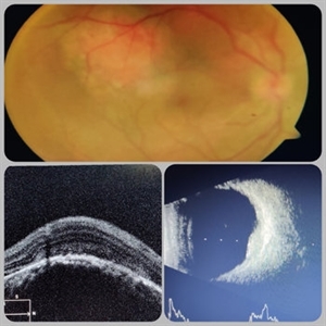 Circumscribed Choroidal Hemangioma
Jul 3 2020 by Dhaivat Shah
A 30-year-old young male presented with drop in vision in right eye since 1 year (6/60). Fundus examination revealed choroidal hemangioma superotemporal to macula. Choroidal hemangioma is an unusual benign vascular tumor of the choroid. It can be circumscribed solitary or diffuse tumor with the later having other systemic associations. Circumscribed choroidal hemangiomas (CCHs) are usually unilateral, unifocal hamartomatous vascular tumor affecting people in second to fourth decade. It appers as round to oval, orangish-red mass in posterior pole with smooth homogenous surface mostly present in macular and peripapillary area. Hyperopic shift is seen in sub-foveal tumors in contrast to para-foveal ones which are usually asymptomatic or present with metamorphopsia or photopsia and diminished vision secondary to exudative retinal detachment. B-scan shows highly reflective tumor without any shadowing or acoustic solidity with high anterior A scan spike. EDI-OCT here depicts a smooth gently sloping choridal mass with compressed choriocapillaries and enlarged medium and large choroidal vessels. Over a period of time structural abnormalities of the outer retina can be visualised. Ancillary testing using Fluorescein Angiography shows lacy hyper-fluorescence during early arterial phase followed by increased hyper-fluorescence due to progressive profuse leakage from pin point foci during arterial and venous phase. Indocyanine green angiography shows lacy diffuse fluorescent tumor in early phase followed by hypo-fluorescent tumor due to dye wash out in late phase. Intrinsic auto-fluorescence is also seen in CCHs from lipofuscin and fresh sub-retinal fluid. Tumor is relatively hyper-intense with respect to vitreous in T1-weighted images in iso-intense in T2-weighted images of MRI. Asymptomatic cases need no treatment, while patients showing vision loss with presence or absence of exudative retinal detachment can be treated with photodynamic therapy which is preferred treatment due to site specific action. Selective occlusion of choroidal neovascularization can be achieved while the neurosensory retinal layers and Bruch membrane are almost unaffected, leaving retinal function intact. Green or rarely red wavelength laser photocoagulation is used to create a chorioretinal adhesion and resolve the SRF. Other treatment modalities include Transpupilary thermotherapy, external beam irradiation, proton beam therapy, brachytherapy and gamma knife.
Photographer: Miss Deepika Nagle
Imaging device: Zeiss
Condition/keywords: B scan ultrasound, choroidal hemangioma, fundus photograph, optical coherence tomography (OCT), photodynamic therapy
-
 Coats' Disease
Jul 10 2018 by Karen Panzegrau
Ultra-wide field images of a 30-year-old male with Coats' Disease affecting his right eye. Patient had sectoral PRP with significant improvement in lipid after 8 months of being lost to folllow up. Vision has improved beyond expectations given severity of lipid.
Photographer: Karen Panzegrau
Imaging device: Optos
Condition/keywords: Coats' disease, fundus photograph, lipid exudation, pan-retinal photocoagulation (PRP)
-
 Coloboma, In Stereo
Oct 1 2012 by Michael P. Kelly, FOPS
This is a stereo retinal fundus photograph of a coloboma, with the optic nerve centered, using a Zeiss FF3C retinal fundus camera.
Photographer: Michael P. Kelly, FOPS Director, Duke Eye Labs, Duke University Hospital, Duke Eye Center, Durham, NC
Condition/keywords: coloboma, fundus photograph, stereo pair
-
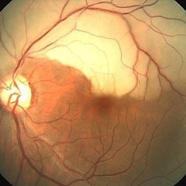
 Combined Hamartoma of the Retina and Retinal Pigment Epithelium (CHRRPE)
Jan 21 2020 by Pierre-Henry Gabrielle, MD
Fundus photograph of a 17-year-old man with combined hamartomas of the retina and retinal pigment epithelium (CHRRPE) at the posterior pole of the left eye.
Photographer: Pierre-Henry Gabrielle, Ophthalmology department, Dijon University Hospital, France
Imaging device: Zeiss Visucam
Condition/keywords: combined hamartoma, fundus photograph
 Loading…