Search results (466 results)
-
 Acute Macular Neuroretinopathy
Sep 15 2014 by Thomas A. Ciulla, MD, MBA, FASRS
Color photo. This might be a typical fundus photo, with no definite lesion. However, the infrared photo nicely depicts a typical lesion.
Condition/keywords: acute macular neuroretinopathy, color photo
-
---thumb.jpg/image-square;max$300,300.ImageHandler) Acute Toxoplasmosis
Acute Toxoplasmosis
Aug 14 2013 by From the Collections of Thomas M. Aaberg, MD and Thomas M. Aaberg Jr., MD
Color and FA sequence.
Condition/keywords: color photo, toxoplasmosis
-
---thumb.jpg/image-square;max$300,300.ImageHandler) Acute Toxoplasmosis
Acute Toxoplasmosis
Aug 14 2013 by From the Collections of Thomas M. Aaberg, MD and Thomas M. Aaberg Jr., MD
Color and FA sequence.
Condition/keywords: color photo, toxoplasmosis
-
---thumb.jpg/image-square;max$300,300.ImageHandler) Acute Toxoplasmosis
Acute Toxoplasmosis
Aug 14 2013 by From the Collections of Thomas M. Aaberg, MD and Thomas M. Aaberg Jr., MD
Color and FA sequence.
Condition/keywords: color photo, toxoplasmosis
-
---thumb.jpg/image-square;max$300,300.ImageHandler) Acute Toxoplasmosis
Acute Toxoplasmosis
Aug 14 2013 by From the Collections of Thomas M. Aaberg, MD and Thomas M. Aaberg Jr., MD
Color and FA sequence.
Condition/keywords: color photo, toxoplasmosis retinitis
-
---thumb.jpg/image-square;max$300,300.ImageHandler) Bergmeister's Papilla
Bergmeister's Papilla
Mar 22 2014 by Hamid Ahmadieh, MD
Color fundus photograph of the right eye of a 50-year-old man with Bergmeister's papilla.
Photographer: Naghmeh Nozhat, Negah Eye Center, Tehran
Imaging device: Topcon Fundus Camera
Condition/keywords: Bergmeister's Papillae, color photo
-
 BRVO Color Photo
Feb 19 2015 by H. Michael Lambert, MD
Superotemporal BRVO.
Condition/keywords: branch retinal vein occlusion (BRVO), color photo
-
---thumb.jpg/image-square;max$300,300.ImageHandler) Butterfly Dystrophy
Butterfly Dystrophy
Aug 12 2013 by From the Collections of Thomas M. Aaberg, MD and Thomas M. Aaberg Jr., MD
Butterfly dystrophy, right eye, color photo.
Condition/keywords: butterfly dystrophy, color photo
-
 Chorioretinal Scar
May 16 2017 by Olivia Rainey
Fundus photograph of an 17-year-old male with a macular scar affecting his right eye secondary to exudation from Coats disease.
Photographer: Olivia Rainey
Imaging device: Topcon 50dx
Condition/keywords: 20 degrees, chorioretinal scar, Coats' disease, color fundus photograph, color photo, fundus photograph
-
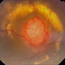
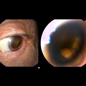 Choroidal Melanoma through the Pupil
May 28 2016 by Olivia Rainey
External image of the left eye of a man with metastatic choroidal melanoma, secondary to lung cancer. There was an obstruction of view to the inferior retina, and this prompted the photographer to pull back to see what the problem was.
Photographer: Olivia Rainey
Imaging device: Topcon 50dx
Condition/keywords: choroidal metastasis, color photo, external photography
-
 Choroidal Melanoma, Color Fundus Photo
Oct 26 2017 by James B. Soque, CRA, OCT-C, COA, FOPS
71-year-old white male with VA 20/40 OD, c/o shadow in upper right quadrant of visual field. Color Montage of right eye reveals choroidal Melanoma. Patient being evaluated at Sloan Kettering, NYC.
Photographer: James B Soque, CRA, OCT-C, COA, FOPS, Island Retina, Shirley, New York
Imaging device: Topcon TRC 50 DX, with MERGE Winstation 11.2.0
Condition/keywords: color fundus photograph, color photo, fundus autofluorescence (FAF), montage
-
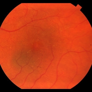 Choroidal Nevus
May 27 2016 by Olivia Rainey
Color fundus image of a small choroidal nevus near the macula.
Photographer: Olivia Rainey
Imaging device: Topcon50dx
Condition/keywords: 20 degrees, choroidal nevus, color fundus photograph, color photo, macula
-
---thumb.jpg/image-square;max$300,300.ImageHandler) Chronic Central Serous Chorioretinopathy
Chronic Central Serous Chorioretinopathy
Feb 20 2013 by From the Collections of Thomas M. Aaberg, MD and Thomas M. Aaberg Jr., MD
Stereo pair color fundus photo of OD of probable bilateral chronic CSR at the macula.
Condition/keywords: chronic central serous chorioretinopathy (CSCR), color photo, stereo pair
-
---thumb.jpg/image-square;max$300,300.ImageHandler) Chronic Central Serous Chorioretinopathy
Chronic Central Serous Chorioretinopathy
Feb 20 2013 by From the Collections of Thomas M. Aaberg, MD and Thomas M. Aaberg Jr., MD
Stereo pair color fundus photo of OS of probable bilateral chronic CSR at the macula.
Condition/keywords: color photo, stereo pair
-
---thumb.jpg/image-square;max$300,300.ImageHandler) Chronic Central Serous Chorioretinopathy
Chronic Central Serous Chorioretinopathy
Feb 20 2013 by From the Collections of Thomas M. Aaberg, MD and Thomas M. Aaberg Jr., MD
Stereo pair color fundus photo of OS of probable bilateral chronic CSR at the macula.
Condition/keywords: chronic central serous chorioretinopathy (CSCR), color photo, stereo pair
-
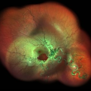 Color Montage Picture
Jan 15 2019 by Aniruddha Maiti, MBBS DO DNB FRVS FICO MRCS FACS FASRS FRCOphth
Dilated tortuous vessels with intraretinal and subretinal hemorrhage at fovea.
Photographer: Sangeeta Mohanta
Imaging device: Heidelberg spectralis
Condition/keywords: color photo, dilated tortuous vessels, subretinal hemorrhage
-
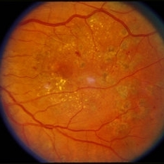
 Color Photo of CSDME
Feb 19 2015 by H. Michael Lambert, MD
Color photo of CSDME.
Condition/keywords: clinically significant macular edema (CSME), color photo, diabetic retinopathy, FALP
-
 Color Photo of CSDME
Feb 19 2015 by H. Michael Lambert, MD
Color photo of CSDME after FALP.
Condition/keywords: clinically significant macular edema (CSME), color photo, diabetic retinopathy, FALP, laser
-
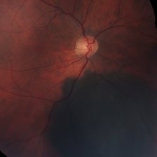
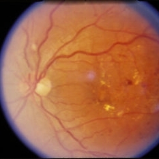
 Color Photo of Optic Disc Capillary Hemangioblastoma
Mar 18 2014 by Arwa Azmeh, MD, PhD
Color fundus photograph of an 48-year-old male who complained of decreased visual acuity in his right eye over the last few months. Systemically the patient was healthy. His VA was OD Cf 3m, OS 20/20. Anterior segments were WNL in OU. IOP was WNL in OU. Fundus exam OD revealed unpigmented mass over the optic disc with retinal venous tortuosity at its edges with a ring of thick HYE surrounding it and shallow RD in this area extending to the foveal area. Several few small retinal hemorrhages were seen in the far retinal periphery which were explained to be caused by venous stasis due the optic disc tumor.
Condition/keywords: color photo, optic disc, retinal hemangioblastoma
-
---thumb.jpg/image-square;max$300,300.ImageHandler) Cone Dystrophy
Cone Dystrophy
Feb 20 2013 by From the Collections of Thomas M. Aaberg, MD and Thomas M. Aaberg Jr., MD
High mag color photo of the macula of OD in a patient with cone dystrophy; VA=20/80.
Condition/keywords: color photo, cone dystrophy, macula
-
---thumb.jpg/image-square;max$300,300.ImageHandler) Cone Dystrophy
Cone Dystrophy
Feb 20 2013 by From the Collections of Thomas M. Aaberg, MD and Thomas M. Aaberg Jr., MD
Color photo of the fundus of OD in a patient with cone dystrophy; VA=20/80.
Condition/keywords: color photo, cone dystrophy, macula
-
---thumb.jpg/image-square;max$300,300.ImageHandler) Cone Dystrophy
Cone Dystrophy
Feb 20 2013 by From the Collections of Thomas M. Aaberg, MD and Thomas M. Aaberg Jr., MD
Green filter photo of the macula of an eye with cone dystrophy.
Condition/keywords: color photo, cone dystrophy, macula
 Loading…