Search results (544 results)
-
 Acute Idiopathic Occlusive Retinal Vasculitis
May 31 2014 by Hamid Ahmadieh, MD
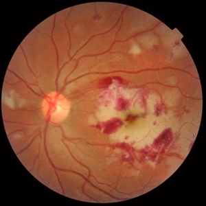
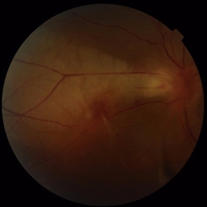
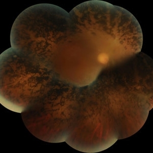
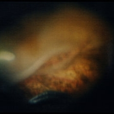
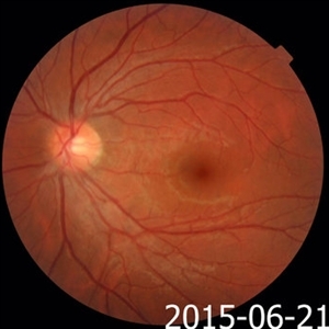
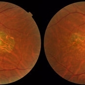
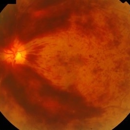
Color fundus photograph of the right eye of a 28-year-old woman with sudden drop of vision due to acute occlusive retinal vasculitis leading to extensive nerve fiber layer infarction and retinal hemorrhages.
Photographer: Naghmeh Nozhat, Negah Eye Center, Tehran
Condition/keywords: color fundus photograph, cotton wool spots, retinal hemorrhage, retinal ischemia
-
 Acute Idiopathic Occlusive Retinal Vasculitis
May 31 2014 by Hamid Ahmadieh, MD
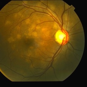
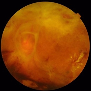
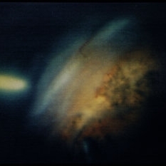
Color fundus photograph of the left eye of a 28-year-old woman with acute drop of vision due to occlusive retinal vasculitis leading to extensive nerve fiber layer infarction and retinal hemorrhages.
Photographer: Naghmeh Nozhat, Negah Eye Center, Tehran
Condition/keywords: color fundus photograph, cotton wool spots, retinal hemorrhage, retinal ischemia
-
 Acute Multifocal Placoid Pigment Epitheliopathy
Sep 15 2014 by Thomas A. Ciulla, MD, MBA, FASRS
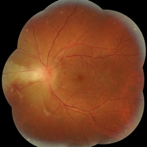
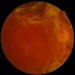
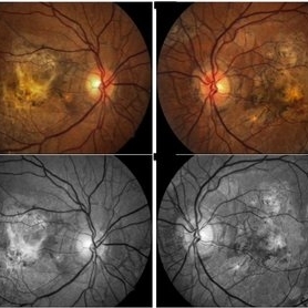
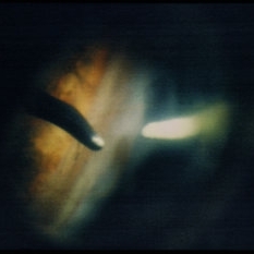
AMPPE in a 42-year-old woman. Color fundus photography shows multiple creamy white subretinal lesions in the superior macula and peripapillary region of the right eye.
Photographer: Thomas Steele
Condition/keywords: acute multifocal placoid pigment epitheliopathy (AMPPE), color fundus photograph
-
 Acute Retinal Necrosis secondary to Herpes Zoster Ophthalmicus
Jan 9 2018 by Olivia Rainey
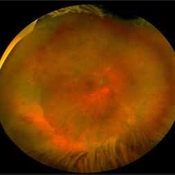
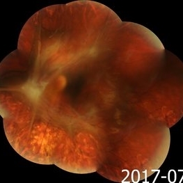
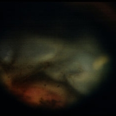
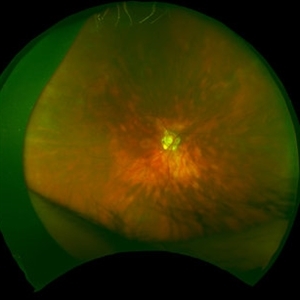
Ultra-wide field Optos pseudocolor montage of an 40-year-old female presenting with acute retinal necrosis secondary to herpes zoster ophthalmicus affecting her right eye.
Photographer: Olivia Rainey
Imaging device: Optos California
Condition/keywords: acute retinal necrosis, color fundus photograph, Herpes zoster, montage, Optos, ultra-wide field imaging
-
 Acute Toxoplasmosis Neuroretinitis
Mar 15 2017 by Hamid Ahmadieh, MD
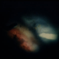
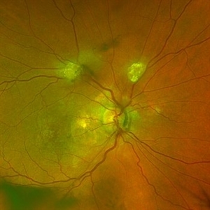
Color fundus photograph of the left eye of a 26-year-old man with clinical picture of acute neuroretinitis and serologic evidence of Toxoplasma gondii infection. Disc swelling, necrotizing retinitis with overlying vitreous inflammation, retinal vasculitis and localized exudative retinal detachment are visible.
Photographer: Solmaz Shahmohammad, Negah Eye Center, Tehran,Iran
Condition/keywords: color fundus photograph, exudative retinal detachment, neuroretinitis, retinal vasculitis, toxoplasmosis
-
 Acute Toxoplasmosis Neuroretinitis
Mar 15 2017 by Hamid Ahmadieh, MD
Color fundus photograph of the left eye of a 26-year-old man with clinical picture of acute neuroretinitis and serologic evidence of Toxoplasma gondii infection. Disc swelling, necrotizing retinitis with overlying vitreous inflammation, retinal vasculitis and localized exudative retinal detachment are visible.
Photographer: Solmaz Shahmohammad, Negah Eye Center, Tehran,Iran
Condition/keywords: color fundus photograph, exudative retinal detachment, neuroretinitis, retinal vasculitis
-
 Advanced PDR
Sep 1 2014 by Hamid Ahmadieh, MD
Color fundus photograph of the left eye of a 50-year-old woman with advanced PDR.
Photographer: Soodabeh Fooladian, Negah Eye Center, Tehran, Iran
Condition/keywords: color fundus photograph, proliferative diabetic retinopathy (PDR)
-
 Advanced PDR
Sep 1 2014 by Hamid Ahmadieh, MD
Color fundus photograph of the right eye of a 50-year-old woman with advanced PDR.
Photographer: Soodabeh Fooladian, Negah Eye Center, Tehran, Iran
Condition/keywords: color fundus photograph, proliferative diabetic retinopathy (PDR), subhyaloid hemorrhage
-
 Advanced Proliferative Diabetic Retinopathy
Nov 4 2017 by Hamid Ahmadieh, MD
Merged color fundus photograph of the left eye of a 30-year-old woman with type1 diabetes since childhood. Note laser scars, severe fibrous proliferation, traction RD and macular dragging.
Photographer: Shabnam Poureh, Negah Eye Center, Tehran, Iran
Condition/keywords: color fundus photograph, diabetes, fibrous proliferation, proliferative diabetic retinopathy (PDR), severe traction
-
 Advanced Retinitis Pigmentosa
Mar 14 2017 by Hamid Ahmadieh, MD
Merged color fundus photograph of the right eye of a patient with advanced retinitis pigmentosa sparing the posterior pole.
Photographer: Soodabeh Fouladin, Negah Eye Center, Tehran, Iran
Condition/keywords: color fundus photograph, retinitis pigmentosa (RP) dystrophy
-
 Angioid Streaks
May 11 2016 by Andrea Arriola-Lopez, MD MSc
64-year-old man, VA CF AO. Inactive neovascularization. Color fundus and red free photograph.
Photographer: Andrea E. Arriola-Lopez MD MSc
Imaging device: Visucam lite Zeiss
Condition/keywords: angioid streaks, color fundus photograph, neovascularization (NV), red-free
-
 Anterior Loop Contraction
Dec 18 2014 by H. Michael Lambert, MD
Anterior loop contraction - intraoperative image.
Condition/keywords: anterior loop contraction, color fundus photograph
-
 Anterior Loop Contraction
Dec 18 2014 by H. Michael Lambert, MD
Anterior loop contraction being addressed with lighted pick and forceps during surgery.
Condition/keywords: anterior loop contraction, color fundus photograph
-
 Anterior Loop Contraction
Dec 18 2014 by H. Michael Lambert, MD
Anterior loop contraction being addressed with lighted pick and forceps during surgery with partial release of traction.
Condition/keywords: anterior loop contraction, color fundus photograph
-
 Anterior Loop Contraction
Dec 18 2014 by H. Michael Lambert, MD
Anterior loop contraction being addressed with lighted pick and forceps during surgery with release of traction.
Condition/keywords: anterior loop contraction, color fundus photograph
-
 Anterior Loop Contraction
Dec 18 2014 by H. Michael Lambert, MD
Anterior loop contraction being addressed with lighted pick and forceps during surgery with release of traction.
Condition/keywords: anterior loop contraction, color fundus photograph
-
 Astrocytic Hamartoma
Feb 27 2025 by Daniel Davis, OCT-C
Color fundus photo of 55-year-old female with Astrocytic Hamartoma in association with tuberous sclerosis. No treatment options available, benign. Other findings include; Posterior Vitreous Detachment, Vitreous Hemorrhage, Hereditary Retinal Dystrophy, Vitreous Opacities, Hypertensive Retinopathy.
Photographer: Daniel Davis, OCT-C
Imaging device: Optos California
Condition/keywords: color fundus photograph
-
 Asymptomatic Eye in FEVR
Jul 7 2015 by Hamid Ahmadieh, MD
Color fundus photograph of the asymptomatic eye of a patient with FEVR. Notice straightening of the retinal vessels.
Photographer: Soulmaz Shahmohammad, Negah Eye Center, Tehran, Iran
Condition/keywords: color fundus photograph, familial exudative vitreoretinopathy (FEVR)
-
 Best Disease
Oct 10 2015 by Hamid Ahmadieh, MD
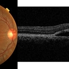
Color fundus photograph and OCT image of the right eye of a 35-year-old man with decreased vision due to Best disease.
Photographer: Shabnam Pooreh, Negah Eye Center, Tehran, Iran
Condition/keywords: Best disease, color fundus photograph, optical coherence tomography (OCT), vitelliform lesion
-
 Best Disease
Oct 10 2015 by Hamid Ahmadieh, MD
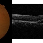
Color fundus photograph and OCT image of the left eye of a 35-year-old man with decreased vision due to Best disease.
Photographer: Shabnam Pooreh, Negah Eye Center, Tehran, Iran
Condition/keywords: Best disease, color fundus photograph, optical coherence tomography (OCT), vitelliform lesion
-
 Birdshot Retinopathy 6
Jan 15 2016 by Raj K. Maturi, MD
Fundus image of an 64-year-old white male.
Photographer: Tom Steele, CRA Midwest Eye Institute Indianapolis, Indiana
Imaging device: Optos
Condition/keywords: birdshot retinochoroidopathy, color fundus photograph
-
 Central Areolar Choroidal Dystrophy
Jul 7 2015 by Hamid Ahmadieh, MD
Color fundus photograph of both eyes of a 58-year-old man with progressive loss of vision. VA OD is 20/60 and VA OS is 20/400.
Photographer: Soulmaz Shahmohammad, Negah Eye Center, Tehran, Iran
Imaging device: Topcon
Condition/keywords: central areolar choroidal dystrophy (CACD), color fundus photograph
-
 Central Retinal Vein Occlusion
Oct 7 2015 by Avris Romario Diparaja Siahaan
Fundus photograph of a 46-year-old man with a central retinal vein occlusion in his left eye.
Photographer: Prayid Listianto, Klinik Mata Nusantara
Imaging device: Topcon TRC 50DX IA
Condition/keywords: central retinal vein occlusion (CRVO), color fundus photograph
-
 Central Retinal Vein Occlusion: Case 1
Oct 12 2012 by Gregg T. Kokame, MD, MMM, FASRS
Color Fundus Photograph
Photographer: Jaclyn Pisano, Retina Consultants of Hawaii
Imaging device: Topcon 50IA / Eschalon
Condition/keywords: central retinal vein occlusion (CRVO)
-
 Central Retinal Vein Occlusion: Case 1
Oct 12 2012 by Gregg T. Kokame, MD, MMM, FASRS
Color Fundus Photograph
Photographer: Jaclyn Pisano, Retina Consultants of Hawaii
Imaging device: Topcon 50IA / Eschalon
Condition/keywords: central retinal vein occlusion (CRVO)
 Loading…