Search results (32 results)
-
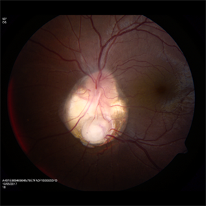
 Cat Eye Syndrome
Feb 11 2020 by Sophia El Hamichi, MD
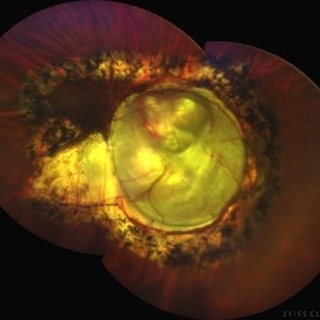
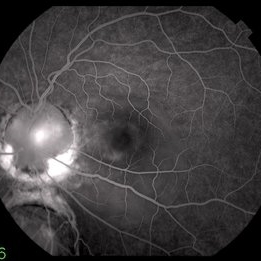
A 3-year-old female with cat eye syndrome including iris, chorioretinal and optic nerve colobomas. Note the CNV temporally to the optic nerve coloboma (blue arrows)
Photographer: Giselle De Oliveira, Bascom Palmer Eye Institute, Miami
Imaging device: RetCam
Condition/keywords: cat eye syndrome, chorioretinal coloboma, choroidal neovascularization (CNV), coloboma, coloboma of optic disc, optic nerve coloboma
-
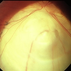
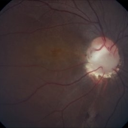
 Chorioretinal coloboma involving disc and macula
Mar 21 2022 by T. P . VIGNESH, MBBS,MS
Fundus photo of Right eye of a 55 year male patient revealing a fovea sparing well barraged chorioretinal coloboma involving the disc and the macula .
Photographer: Bharathi Singaravel
Imaging device: Zeiss Clarus
Condition/keywords: chorioretinal coloboma, coloboma of optic disc
-
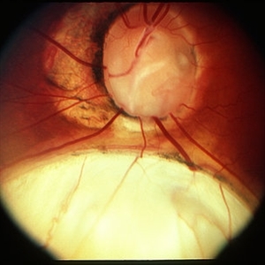
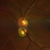
 Coloboma
Mar 29 2013 by Henry J. Kaplan, MD
Coloboma involving optic nerve and inferior choroid.
Condition/keywords: coloboma of choroid, coloboma of optic disc
-
 Coloboma
Mar 29 2013 by Henry J. Kaplan, MD
Optic disc and inferonasal choroidal coloboma in the same patient #2.
Condition/keywords: coloboma, coloboma of choroid, coloboma of optic disc
-
 Coloboma
Sep 17 2015 by Jason S. Calhoun
Fundus photograph of young female with retinal coloboma.
Photographer: Jason Calhoun, Mayo Clinic, Department of Ophthalmology
Condition/keywords: coloboma of optic disc
-
 Coloboma involving the Optic nerve, Retina, and Choroid
Dec 6 2021 by Jesus Lozano, MD
78-year-old woman after prophylactic laser photocoagulation (PLP) for her RE Coloboma involving the optic nerve, retina, and choroid. At 6 month follow up, patient preserved her FC vision as it was before the procedure. Retina attached.
Photographer: Yair Bet Yosef, Hadassah Medical Center. Israel
Imaging device: Optos Silverstone fundus image
Condition/keywords: coloboma, coloboma of choroid, coloboma of macula, coloboma of optic disc, PLP, prophylactic photocoagulation
-
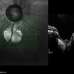
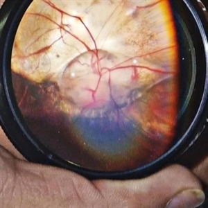
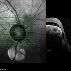 Coloboma of Disc & Choroid
Oct 6 2012 by Hamid Ahmadieh, MD
OCT image of a 25-year-old woman with serous retinal detachment secondary to coloboma of disc associated with coloboma of choroid.
Photographer: Hamid Ahmadieh, MD, Ophthalmic Research Center, Labbafinejad Medical Center, Shahid Beheshti University of Medical Sciences
Imaging device: Heidelberg Spectralis
Condition/keywords: coloboma of choroid, coloboma of optic disc, optical coherence tomography (OCT), serous retinal detachment
-
 Coloboma of Disc & Choroid
Oct 6 2012 by Hamid Ahmadieh, MD
OCT image of a 25-year-old woman with a small coloboma of choroid associated with coloboma of disc.
Photographer: Hamid Ahmadieh, MD, Ophthalmic Research Center, Labbafinejad Medical Center, Shahid Beheshti University of Medical Sciences
Imaging device: Heidelberg Spectralis
Condition/keywords: coloboma of choroid, coloboma of optic disc, optical coherence tomography (OCT)
-
 Coloboma of Optic Disc
Apr 28 2019 by Bastián Schmidt Arias
Fundus photograph of an 63-year-old woman with retinal coloboma.
Photographer: Bastian Schmidt
Condition/keywords: coloboma of optic disc
-
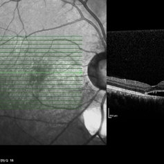
 Coloboma of Optic Disc
Sep 23 2022 by Kavya Rao, M.S
OCT and OCT Angiography (4.5x4.5mm)(ZEISS) of 39-year old man ,came for routine check up and diagnosed with coloboma of Optic Disc in the Right Eye as an incidental finding.
Photographer: Dr.KAVYA RAO, LIONS CLUB OF HYDERABAD, SADHURAM EYE HOSPITAL,HYDERABAD,INDIA
Condition/keywords: coloboma
-
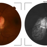
 Colobomatous Optic Disc Maculopathy
Feb 13 2020 by Yoshihiro Yonekawa, MD, FASRS
Beautifully focused fundus photograph of a teenage girl with submacular fluid from a colobomatous optic disc.
Photographer: Netanya Lerner, COA, Wills Eye Hospital/Mid Atlantic Retina
Imaging device: Topcon
Condition/keywords: chorioretinal coloboma, coloboma of optic disc, congenital optic nerve pit, subretinal fluid
-
 Colobomatous Optic Disc Maculopathy
Feb 13 2020 by Yoshihiro Yonekawa, MD, FASRS
Fluorescein angiography, late frame, of a teenage girl with submacular fluid from a colobomatous optic disc. The camera is focused is on the elevated macula, and the disc is subtly defocused.
Photographer: Netanya Lerner, COA, Wills Eye Hospital/Mid Atlantic Retina
Imaging device: Topcon
Condition/keywords: chorioretinal coloboma, coloboma of optic disc, congenital optic nerve pit, subretinal fluid
-
 Colobomatous Optic Disc Maculopathy
Feb 13 2020 by Yoshihiro Yonekawa, MD, FASRS
EDI-OCT of a teenage girl with submacular fluid from a colobomatous optic disc. Note the subtle tracking of the subretinal fluid into the disc.
Photographer: Netanya Lerner, COA, Wills Eye Hospital/Mid Atlantic Retina
Imaging device: Topcon
Condition/keywords: chorioretinal coloboma, coloboma of optic disc, congenital optic nerve pit, subretinal fluid
-
 Disc Coloboma
Aug 17 2023 by Dr.Anushri Godbole
21 years old female came to OPD with chief complaints of diminution of vision of LE since birth. BCVA RE-6/6 N6, LE FC-1/2M, N36. On examination RE was diagnosed as disc coloboma with type 6 coloboma in periphery and LE was diagnosed as Choroidal coloboma involving disc
Condition/keywords: coloboma of choroid, coloboma of optic disc
-
 Fundal Coloboma
Sep 25 2024 by DR Rohit Gupta
Fundus photograph of 16year old female patient with a fundal coloboma in left eye
Photographer: Dr Rohit gupta
Imaging device: Samsung S21
Condition/keywords: chorioretinal coloboma, coloboma of macula, coloboma of optic disc, congenital anomaly
-
 Fundus Coloboma
Feb 22 2023 by Zach Seim
An ultra-widefield fundus image of a 25 year old male with Fundus Coloboma, as well as Iris Coloboma affecting both eyes. Patient's vision at the time of the image was 20/100-2. Discussed genetic testing as patient reports that he has a child with coloboma and patient agrees. There is a possibility of this finding being syndromic given cornea has small WTW and possibly microphthalmia. The patient has old tractional exudation at edge (abutting fovea). Recommended observation without treatment.
Photographer: Zach Seim
Imaging device: Optos California
Condition/keywords: coloboma, coloboma of optic disc, fundus photograph, Optos, scanning laser ophthalmoscope, ultra-wide field imaging
-
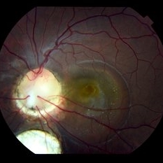
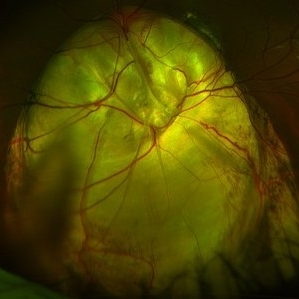
 Morning Glory Disc Anomaly
Aug 19 2017 by Mitzy E Torres Soriano, MD
A 10-year-old female patient with morning glory disc anomaly in her left eye.
Photographer: Mitzy E. Torres Soriano
Condition/keywords: coloboma of optic disc, coloboma of the optic nerve, Morning Glory Syndrome, optic disc, optic disc dysplasia
-
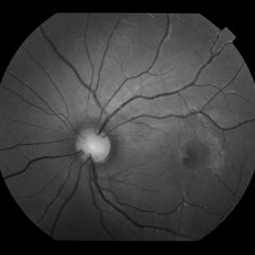
 Optic Disc Coloboma
Aug 27 2022 by Aditya S Kelkar, MS, FRCS, FASRS,FRCOphth
Color fundus photograph of a 51-year-old man showing optic disc coloboma of the left eye.
Photographer: Dr. Sukanya Mondal. National Institute of Ophthalmology, Pune, India.
Imaging device: Zeiss Clarus 500
Condition/keywords: coloboma of optic disc, color fundus photograph
-
 Optic Disc Coloboma
May 12 2017 by Nimrod Dar
9-year-old patient, noticed a gradual deterioration in her visual acuity at her LE (6/15). On her examination, an optic disc coloboma / pit can be seen. OCT scan revealed an intra retinal fluid and maculo schisis
Photographer: Nimrod Dar
Condition/keywords: coloboma, coloboma of optic disc, optic disc
-
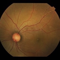
 Optic Disc Coloboma
Oct 17 2018 by Mehul A Shah
14-year-old girl at routine check-up and fundus picture found optic disc coloboma.
Photographer: MEHUL SHAH
Condition/keywords: coloboma of optic disc
-
 Optic Disc Coloboma
Jul 24 2019 by Haider Ali
16-year-old boy with horizontal nystagmus and decreased vision in both eyes.
Photographer: Dr Haider Ali Chaudhry, Madinah Teaching Hospital, Faisalabad
Condition/keywords: coloboma, coloboma of optic disc, coloboma of the optic nerve, excavation, Morning Glory Syndrome
-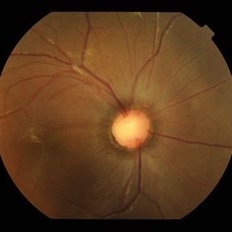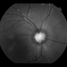
 Optic Disc Coloboma
Jul 24 2019 by Haider Ali
16-year-old boy with horizontal nystagmus and decreased vision in both eyes.
Photographer: Dr Haider Ali Chaudhry, Madinah Teaching Hospital, Faisalabad
Condition/keywords: coloboma, coloboma of optic disc, coloboma of the optic nerve, excavation, Morning Glory Syndrome
 Loading…