Search results (24 results)
-
 Arterial Occlusion
Jul 25 2019 by JEFFERSON R SOUSA, Tecg.º (Biomedical Systems Technology)
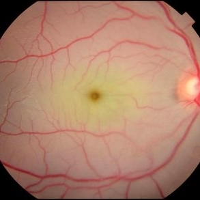
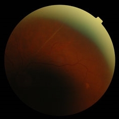
Male patient 16-years-old, was admitted to the clinic with low vision failure. On evaluation, signs of arterial occlusion in the right eye were observed. The imaging exams in the clinical evaluation showed important changes in the blood flow of one of the carotid arteries (partial obstruction), probably atherosclerotic carotid disease.
Photographer: JEFFERSON R SOUSA - Study Center and Ophthalmological Research Dr. Andre M V Gomes, Institute Dr. Suel Abujamra São Paulo-Brazil
Imaging device: Topcon TRC-50 DX, Imaginet 5.0, angle de 50 graus. Flash 36
Condition/keywords: arterial occlusion
-
 Arterial Occlusion
Jul 14 2013 by Jason S. Calhoun
Patient in with vision loss in the lower quadrant of his visual field. Fundus photo shows arterial occlusion superior to the optic nerve
Photographer: Jason S. Calhoun, Department of Ophthalmology, Mayo Clinic Jacksonville, Florida
Imaging device: TOPCON TRC 50-EX
Condition/keywords: branch retinal artery occlusion (BRAO)
-
 Arterial Occlusion With Preservation of Cilioretinal Network
Aug 29 2016 by JEFFERSON R SOUSA, Tecg.º (Biomedical Systems Technology)
Patient male, 52-years-old, denies hypertension, blood systemic, and diabetes. Refers low subtle of vision, field loss and stain central.
Photographer: JEFFERSON R SOUSA - Institute Dr. Suel Abujamra / São Paulo - Brazil
Imaging device: Acquisition of the image in the Camera background Topcon TRC-50 Dx - IA, Keystone field photo of 50 Degrees. Composition automatic of Imaginet with manual adjustment.
Condition/keywords: arterial occlusion, hypertension
-
 BRAO (Branch Retinal Artery Occlusion)
Jun 24 2023 by Mauricio Galvan Chavez, MD
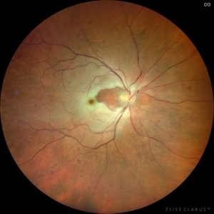
Fundus photograph of a BRAO (Branch Retinal Artery Occlusion)
Photographer: Mauricio Galvan, Clinica de Retina, Guadalajara Jalisco.
Imaging device: Zeiss Clarus 700
Condition/keywords: arterial occlusion, branch retinal artery occlusion (BRAO), BRAO
-
 Cilioretinal artery occlusion
Mar 12 2023 by Pawel Kolman
58 year-old male with sudden, painless vision loss of RE. In doppler ultrasound there was found 90% arterioscleral stenosis of right internal carotid artery.
Photographer: Pawel Kolman
Imaging device: Volk 20D and Samsung Galaxy S21
Condition/keywords: arterial occlusion, cilioretinal artery occlusion
-
---thumb.jpg/image-square;max$300,300.ImageHandler) Cilioretinal Artery Occlusion
Cilioretinal Artery Occlusion
Oct 18 2012 by Larry Halperin, MD
Cilioretinal artery occlusion
Condition/keywords: arterial occlusion, cilioretinal artery occlusion
-
 Mixed Occlusion of Artery and Vein
Jan 6 2021 by Renata Garcia Franco, Md
Male with a history of smoking, sudden low vision of the right eye, retinal neovascularization and inferior preretinal hemorrhage.
Photographer: Fatima Hernandez, Instituto de la Retina del Bajio SC
Imaging device: Zeiss
Condition/keywords: arterial occlusion
-
 Ophthalmic Artery Occlusion
Jan 28 2017 by ADRIANO FERREIRA
32-year-old female with a sudden loss of vision in the left eye. This fundus photograph shows central arterial occlusion plus central vein occlusion secondary to ophthalmic artery occlusion. This patient has thromboangitis obliterans.
Photographer: Mr. Jose Luiz
Imaging device: TRC50DXi
Condition/keywords: arterial occlusion, occlusion of retinal vein
-
 Superior Hemi-Central Retinal Artery Occlusion
Apr 24 2024 by Mosab Salah
Fundus photograph -inverted view- taken by smartphone fundus photography, of a young man with sudden onset altitudinal field defect, a Superior Hemi-Central Retinal Artery Occlusion noted.
Photographer: Dr Mosab Salah, The Islamic Hospital, Amman, Jordan
Imaging device: smartphone fundus photography and 30 D Lens
Condition/keywords: arterial occlusion, branch retinal artery occlusion (BRAO), BRAO, CRAO, Hemi-Central Retinal Artery Occlusion (CRAO), occlusive vasculitis, smartphone fundus photography
-
 Venous Stasis Retinopathy
Aug 6 2024 by Akansha Sharma
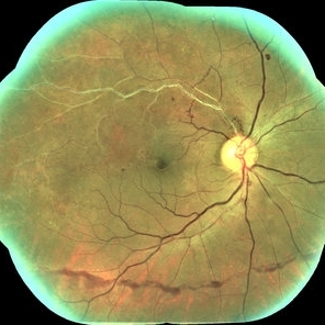
Color fundus photograph of a 18 year old male with venous stasis retinopathy.
Photographer: Dr. Akansha Sharma, Bharati Eye Hospital
Condition/keywords: arterial occlusion, venous stasis retinopathy
-
 Venous Stasis Retinopathy
Feb 20 2015 by H. Michael Lambert, MD
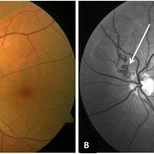
Scattered hemorrhages associated with venous stasis retinopathy in eye with ipsilateral total internal carotid artery occlusion. Vision is 20/40.
Condition/keywords: arterial occlusion, venous stasis retinopathy
-
 Venous Stasis Retinopathy
Feb 20 2015 by H. Michael Lambert, MD
Scattered hemorrhages associated with venous stasis retinopathy in eye with ipsilateral total internal carotid artery occlusion. Vision is 20/40.
Condition/keywords: arterial occlusion, venous stasis retinopathy
-
 Venous Stasis Retinopathy
Feb 20 2015 by H. Michael Lambert, MD
68 second fluorescein angiogram of eye with venous stasis retinopathy in eye with ipsilateral total internal carotid artery occlusion. Fluorescein angiogram shows slow fill. Vision is 20/40.
Condition/keywords: arterial occlusion, venous stasis retinopathy
-
 Venous Stasis Retinopathy
Feb 20 2015 by H. Michael Lambert, MD
53 second fluorescein angiogram of eye with venous stasis retinopathy in eye with ipsilateral total internal carotid artery occlusion. Fluorescein angiogram shows slow fill. Vision is 20/40.
Condition/keywords: arterial occlusion, venous stasis retinopathy
-
 Venous Stasis Retinopathy
Feb 20 2015 by H. Michael Lambert, MD
Scattered hemorrhages associated with venous stasis retinopathy in eye with ipsilateral total internal carotid artery occlusion. Vision is 20/40.
Condition/keywords: arterial occlusion, venous stasis retinopathy
-
 Branch Arterial Occlusion
Apr 12 2018 by Marco D'Angelo
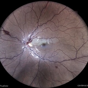
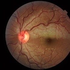
Branch artery occlusion with collateral circulation (arterio-arterial vascular anastomosis) around the residual perfused branch. Left eye, 76-years-old male patient, normal visual acuity (20/20).
Photographer: Dr. Marco D'Angelo, S.Chiara Hospital, Trento, Italy
Imaging device: Topcon TRC-NW 6S
Condition/keywords: arteriovenous anastomosis, collateral retinal vessel, ghost vessels
-
 Branch Retinal Artery Occlusion
May 4 2021 by Priya Rasipuram Chandrasekaran, MBBS, DO, DNB, FRCS
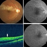
This is the fundus photo of a 52-year-old male taken within 6 hours and after 24 hours of sudden onset of inferior field loss. The photo shows prominence of retinal edema in the region of arterial occlusion as time passes by. The optical coherence tomogram scan taken vertically through the normal and the involved area shows thickening and hyper reflectivity of retinal nerve fiber layer and decreased reflectivity of the retinal layers beneath it (white arrow). Fundus fluorescein angiography shows complete non-filling of the artery in the early phase with slow filling in the late phase and highlighting the embolus.
Condition/keywords: branch retinal artery occlusion (BRAO)
-
 Branch Retinal Artery Occlusion (BRAO)
Sep 12 2023 by Ben Serar
Fundus photograph of the LE showing arterial occlusion along the inferotemporal vessel arcade with surrounding retinal edema and cotton-wool spots, in a case of Branch Retinal Artery Occlusion (BRAO).
Condition/keywords: branch retinal artery occlusion (BRAO), cotton wool spots, retinal edema
-
 Peri-papillary Vascular Loop
Jun 2 2020 by Dhaivat Shah
Peri-papillary vascular loops (PVL) are rare congenital vascular malformations, which are usually detected as accidental finding during routine fundus examination. They can often be confused with tributary vein occlusion or racemose hemangioma. Although benign and asymptomatic, they can be rarely associated with vitreous hemorrhage and arterial occlusion. We herein present a case of a 60-year-old hypertensive male, who was diagnosed elsewhere to have a tributary vein occlusion and was referred to us. FFA was advised to rule out neovascularization, surrounding capillary non perfusion and mass lesion (hemangioma). On FFA, the arterial loop showed a slightly delayed filling (3-5 seconds) as compared to the other arterial vessels and the original vessel appeared to be a branch arising from central retinal artery. The choroidal filling was delayed in the area supplied by the loop. A cilioretinal artery was also noted. The patient was diagnosed to have a Peri-papillary vascular arterial loop (PVL), likely to be congenital in origin. The patient was reassured and was advised yearly follow up. These loops are usually accidental findings discovered during routine fundus examination. Since these vessels are looped and tortuous, they exhibit a slower and laminar blood flow, which make them more prone for arterial occlusions. The vitreous in this area tends to be adherently attached, so during PVD induction, it is likely to cause a tear and hemorrhage leading to vitreous hemorrhage. Until and unless there is a break, this hemorrhage tends to resolve on its own and does not warrant treatment. If there is an evident break, it can be dealt with laser barrage.
Photographer: Choithram Netralaya
Condition/keywords: congenital prepapillary vascular loop
-
 Purtscher's Like Retinopathy
Aug 22 2012 by Ivan R. Batlle, MD
72-year-old man with sudden loss of vision in the left eye shortly after renal artery catheterization.
Condition/keywords: partial arterial occlusion
-
 Purtscher's Like Retinopathy
Aug 22 2012 by Ivan R. Batlle, MD
Red Free image of a 72-year-old man with sudden loss of vision shortly after a renal artery catheterization.
Condition/keywords: partial arterial occlusion
-
 Purtscher's LIke Retinopathy
Aug 22 2012 by Ivan R. Batlle, MD
Mid-phase fluorescein angiogram of a 72-year-old man with sudden loss of vision shortly after a renal artery catheterization.
Condition/keywords: partial arterial occlusion
-
 Purtscher's Like Retinopathy
Aug 22 2012 by Ivan R. Batlle, MD
Late Phase Fluorescein Angiogram of a 72-year-old man with sudden loss of vsion shortly after a renal artery catheterization.
Imaging device: Zeiss
Condition/keywords: partial arterial occlusion
-
 WAXING MOON OR WANING MOON?
Oct 12 2023 by Deepti A Kulkarni, M.B.B.S., D.N.B., F.V.R.
FUNDUS PHOTO OF AN 18 YEAR OLD. VISION AT PRESENTATION 6/6 WITH DIFFICULTY IN READING AND A BLACK SPOT WHEN TRYING TO FOCUS. ANAEMIC FOR OVER TWO YEARS - IN HER RECOVERY PHASE WHERE HEMOGLOBIN HAD RISEN FROM <5g/dL TO 7.5 g/dL WHERE SHE HAD REACTIVE THROMBOCYTOSIS CAUSING HYPERCOAGULABILITY AND RETINAL ARTERIOLAR OCCLUSION.
Photographer: DEEPTI KULKARNI, KULKARNI EYE HOSPITAL, MIRAJ, INDIA
Imaging device: TOPCON
Condition/keywords: ANEMIC RETINOPATHY, MACULAR BRANCH ARTERIAL OCCLUSION
 Loading…