Search results (15 results)
-
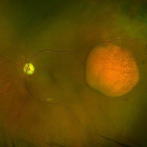
 Amelanotic Choroidal Melanoma
Apr 12 2019 by David L Kilpatrick, MD
Fundus photograph of a 69-year-old male with an amelanotic choroidal melanoma and corresponding exudative retinal detachment. Transvitreal biopsy was performed at the time of radioactive I-125 plaque placement. The genetic expression profile revealed a Class 1A, PRAME negative tumor.
Photographer: Retina Consultants of Alabama, P. C.
Imaging device: Optos
Condition/keywords: amelanotic melanoma
-
 Amelanotic Choroidal Melanoma
May 18 2020 by McGill University Health Centre
The enucleation image shows a large amelanotic tumor with large areas of hemorrhage and necrosis. Note the several dilated blood vessels and an adjacent retinal detachment with lipofuscin pigment on its surface (arrow).
Condition/keywords: amelanotic melanoma, enucleation, mushroom-shaped
-
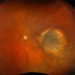
 Amelanotic Choroidal Melanoma with Optic Atrophy
Jun 11 2025 by Aditya S Kelkar, MS, FRCS, FASRS,FRCOphth
Fundus photograph of a 64-year-old woman with optic atrophy and amelanotic choroidal melanoma temporal to the macula.
Photographer: Dr Harsh Jain, National Institute of Ophthalmology
Imaging device: Optos Daytona
Condition/keywords: amelanotic melanoma, optic atrophy
-
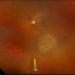
 Amelanotic Melanoma
Oct 5 2015 by Scott C. Oliver, MD
58-year-old female with inferonasal choroidal tumor detected on routine eye exam.
Photographer: William Yates, UCHealth Eye Center
Imaging device: Topcon TRC-50DX
Condition/keywords: melanoma
-
 Amelanotic Melanoma
Aug 12 2025 by César Adrián Gómez Valdivia, MD
This case highlights an amelanotic melanoma, an atypical presentation of a choroidal melanoma lacking the characteristic pigmentation. These lesions can easily be mistaken for choroidal hemangiomas, metastases, or inflammatory masses. Clinically, the lesion appears as a dome-shaped, yellowish subretinal mass, often associated with subretinal fluid, lipofuscin deposition, or retinal detachment. The absence of pigment can delay diagnosis, making multimodal imaging essential. Diagnostic tools: • B-scan ultrasound: low to medium internal reflectivity • OCT: overlying subretinal fluid and RPE elevation • FAF: orange pigment and RPE disruption • ICG/FA: variable, often hypofluorescent core Important: Prompt referral to ocular oncology is critical for management and prognosis.
Photographer: @eyemissu2
Imaging device: TOPCON TRC-50DX
Condition/keywords: amelanotic melanoma
-
 Amelanotic Melanoma
Aug 12 2025 by César Adrián Gómez Valdivia, MD
This FAF image reveals a hypoautofluorescent mass with areas of dense hyperautofluorescent stippling—a classic pattern suggestive of an amelanotic choroidal melanoma. Amelanotic melanoma is a rare variant of uveal melanoma, accounting for only a minority of cases. Unlike pigmented melanomas, these lesions lack melanin, making them more challenging to detect on conventional color fundus imaging. FAF Characteristics: • Central hypoautofluorescence: due to loss or compression of the RPE • Peripheral hyperautofluorescent speckling: consistent with lipofuscin accumulation or RPE disruption • Often associated with subretinal fluid or orange pigment seen clinically Location: Juxtapapillary, with potential optic nerve involvement—a factor that complicates both diagnosis and
Photographer: @eyemissu2
Imaging device: California ICG OPTOS
Condition/keywords: amelanotic melanoma
-
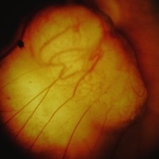
 Amelanotic Mushroom-Shaped Choroidal Melanoma
May 18 2020 by McGill University Health Centre
This enucleation specimen demonstrates an amelanotic, mushroom-shaped, slightly hemorrhagic tumor near the optic nerve (arrow). True retinal detachment is present, and the retina is folded (arrowhead). The subretinal fluid is hazy (*).
Condition/keywords: amelanotic melanoma, enucleation, mushroom-shaped
-
 New Choroidal Melanoma
Jul 16 2025 by Virginia Gebhart
78 year old male with a partially amelanotic dome-shaped lesion with RPE changes, hard exudates, overlying intraretinal fluid and minimal SRF temporally. Exam and ultrasound findings consistent with choroidal melanoma. Pt will be scheduled for brachytherapy pending CT scan results.
Photographer: Virginia Gebhart, Retina Consultants of Carolina
Imaging device: Optos California
Condition/keywords: amelanotic melanoma, choroidal melanoma
-
 New Choroidal Melanoma vs Metastasis
Dec 6 2023 by Virginia Gebhart
72 year old male with possible new choroidal melanoma vs metastatic melanoma. Dome-shaped amelanotic lesion involving the fovea. Lesion was discovered during a problem visit due to sudden decreased VA (20/150). Most recent CT scan shows concern for primary lung cancer
Photographer: Virginia Gebhart
Imaging device: Topcon
Condition/keywords: amelanotic melanoma, melanoma, metastatic lesion
-
 New Iris Melanoma
Oct 10 2024 by Virginia Gebhart
56 year old male with new amelanotic melanoma emanating from the ciliary body through the posterior iris epithelium. CT scan showed no evidence of metastatic disease. Pt scheduled for radioactive plaque and tumor biopsy
Photographer: Virginia Gebhart, Retina Consultants of Carolina
Imaging device: Samsung Galaxy
Condition/keywords: amelanotic melanoma, iris melanoma
-
 Treated Melanoma with Iluvien Implant
Apr 9 2025 by Virginia Gebhart
62 year old female 4 mo s/p brachytherapy for amelanotic choroidal melanoma. Iluvien implant given 4 wks s/p plaque removal, lesion is stable with resolved exudative detachment and subretinal fluid
Photographer: Virginia Gebhart, Retina Consultants of Carolina
Imaging device: Optos California
Condition/keywords: amelanotic melanoma, brachytherapy, choroidal melanoma, Iluvien, melanoma
-
 Juxtapapiillary Choroidal Amelanotic Melanoma - Fundus Photograph
Aug 27 2014 by Roy Schwartz, MD
Fundus photograph of a juxtapapillary choroidal amelanotic melanoma.
Photographer: Daniel Gold, MD
-
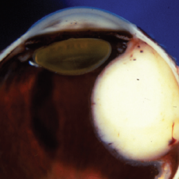
 Juxtapapiillary Choroidal Amelanotic Melanoma - Gross Pathology
Aug 27 2014 by Roy Schwartz, MD
Gross pathology of a juxtapapillary choroidal amelanotic melanoma.
Photographer: Daniel Gold, MD
Condition/keywords: gross pathology, gross specimen
-
 Juxtapapiillary Choroidal Amelanotic Melanoma - Histopathology
Aug 27 2014 by Roy Schwartz, MD
Histopathology of a juxtapapillary choroidal amelanotic melanoma, showing a spindle B melanoma pattern.
Photographer: Daniel Gold, MD
Condition/keywords: histopathology, pathology
-
 Leiomyoma
May 18 2020 by McGill University Health Centre
Leiomyoma is a benign, smooth muscle tumor. Ninety percent of cases occur in women. The differential diagnosis includes amelanotic melanoma and nerve sheath tumors. This transversal pupil–optic nerve (PO) section of an enucleation specimen shows a nodular, well-delineated, whitish tumor in the ciliary body. The cut surface shows small foci of hemorrhage without necrosis. The retina partially covers the inner surface of the tumor, and the sclera is not infiltrated. Note the slightly displaced (subluxated) cataractous lens and the choroidal detachment artifact in the right inferior corner.
Condition/keywords: enucleation, leiomyoma, tumor
 Loading…