Search results (212 results)
-
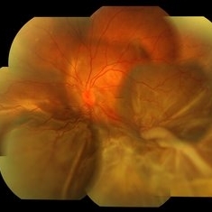
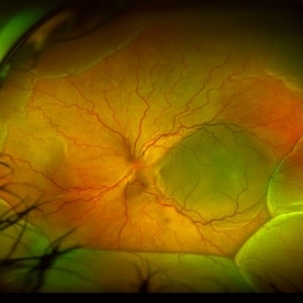
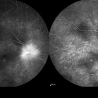
 "Starry Sky" Fundus in Vogt-Koyanaki-Harada Syndrome
Jan 10 2018 by Peter H. Tang, MD, PhD
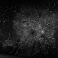
Fluorescein angiography imaging of a 27-year-old male with acute inflammation as part of Vogt-Koyanagi-Harada Syndrome.
Imaging device: Optos California
Condition/keywords: chorioretinal inflammations, retina, uveitis, Vogt-Koyanagi-Harada
-
 2min-FA-VKH
Oct 20 2021 by Bryon R McKay, MD, PhD, FRCSC, DRCPSC - Retina
27yF presented with sub-acute findings of VKH, she has an interesting pattern of perivascular changes. She was successfully treated with immunosuppressive agents and maintains 20/20 vision.
Photographer: Dr. K. Vaezi, University of British Columbia, Canada
Imaging device: Optos Imaging system
Condition/keywords: uveitis, Vogt-Koyanagi-Harada
-
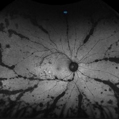 Autofluorescence Stage 3 Vogt-Koyanagi-Harada (VKH) Disease
Oct 20 2021 by Bryon R McKay, MD, PhD, FRCSC, DRCPSC - Retina
27yF presented with sub-acute findings of VKH, she has an interesting pattern of perivascular changes. She was successfully treated with immunosuppressive agents and maintains 20/20 vision.
Photographer: Dr. K. Vaezi, University of British Columbia, Canada
Imaging device: Optos Imaging system
Condition/keywords: Vogt-Koyanagi-Harada
-
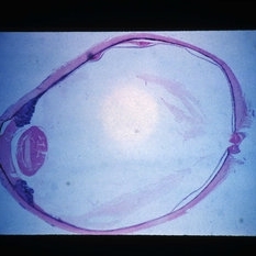
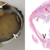 Chronic Vogt-Koyanagu-Harada (VKH) Disease
May 18 2020 by McGill University Health Centre
This enucleation specimen shows complete distortion of the anterior chamber. The iris and the chamber are completely replaced by a whitish mass corresponding to inflammatory exudate. The eye is aphakic and the retina is completely detached from the choroid, although it is attached anteriorly to the mass. Note the proteinaceous exudate beneath the retina (arrowheads).
Condition/keywords: chronic, Vogt-Koyanagi-Harada
-
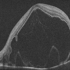 Exudative Detachment of the Macula in Vogt-Koyanagi-Harada Syndrome
Jan 10 2018 by Peter H. Tang, MD, PhD
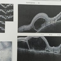
SD-OCT imaging of an exudative detachment of the macula in a 27-year-old male diagnosed with Vogt-Koyanagi-Harada Syndrome.
Imaging device: Zeiss Cirrus HD-OCT
Condition/keywords: exudative macula detachment, serous retinal detachment, uveitis, Vogt-Koyanagi-Harada
-
 Multifocal CSR With Exudative RD
May 19 2017 by Manish Nagpal, MD, FRCS (UK), FASRS
A 30-year-old male diagnosed elsewhere as VKH was started on heavy steroids and he developed multiple serous elevations and OS developed a exudative RD.
Photographer: POOJA BAROT
Condition/keywords: central serous retinopathy (CSR), multifocal central serous chorioretinopathy (CSCR), Vogt-Koyanagi-Harada
-
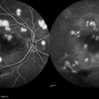 Multifocal CSR FA & ICG
May 19 2017 by Manish Nagpal, MD, FRCS (UK), FASRS
A 30-year-old male diagnosed elsewhere as VKH was started on heavy steroids and he developed multiple serous elevations and OS developed a exudative RD. We immediately asked the patient to stop steroids and when he followed up after a month lesions had flattened and he had recovered to 20/40 in both eyes.. he is still undergoing further follow up at this stage...
Photographer: pooja barot
Imaging device: heidelberg
Condition/keywords: central serous retinopathy (CSR), multifocal central serous chorioretinopathy (CSCR), Vogt-Koyanagi-Harada
-
 Multifocal CSR OD
May 19 2017 by Manish Nagpal, MD, FRCS (UK), FASRS
A 30-year-old male diagnosed elsewhere as VKH was started on heavy steroids and he developed multiple serous elevations and OS developed a exudative RD.
Photographer: POOJA BAROT
Condition/keywords: central serous retinopathy (CSR), multifocal central serous chorioretinopathy (CSCR), Vogt-Koyanagi-Harada
-
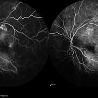 Multifocal CSR OS FA and ICG
May 19 2017 by Manish Nagpal, MD, FRCS (UK), FASRS
A 30-year-old male diagnosed elsewhere as VKH was started on heavy steroids and he developed multiple serous elevations and OS developed a exudative RD.
Photographer: pooja barot
Condition/keywords: central serous retinopathy (CSR), Vogt-Koyanagi-Harada
-
 Optos FA of Harada's Disease
Jun 22 2013 by James A Eadie, MD
Optos fluorescein angiogram of a 22-year-old woman with Harada's disease. An exudative detachment is billowing in the foreground at the bottom of the image.
Photographer: John Peterson
Imaging device: Optos camera
Condition/keywords: Harada's disease, Vogt-Koyanagi-Harada
-
 Recurrent VKH
Mar 8 2022 by mohamed ahmed el shafie, ophthalmic resident
A case of recurrent VKH. OCT scan
Condition/keywords: subretinal fluid, Vogt-Koyanagi-Harada
-
 Resolved Exudative RD in Vogt-Koyanagi-Harada Syndrome
Mar 27 2019 by Gary R. Cook, MD, FACS
Right eye of the same patient following resolution of the exudative retinal detachment OD.
Condition/keywords: exudative detachment, panuveitis, Vogt-Koyanagi-Harada
-
 Serous Retinal Detachment
Oct 14 2024 by César Adrián Gómez Valdivia, MD
Serous Retinal Detachment found in a 19 year-old female patient with suspected Vogt-Koyanagi-Harada disease. Findings were bilateral. Patient was admitted for Methylprednisolone and Cyclophosphamide treatment.
Photographer: @eyemissu2
Imaging device: California ICG OPTOS
Condition/keywords: serous retinal detachment, Vogt-Koyanagi-Harada
-
 Serous Retinal Detachment
Oct 14 2024 by César Adrián Gómez Valdivia, MD
Serous Retinal Detachment found in a 19 year-old female patient with suspected Vogt-Koyanagi-Harada disease. Findings were bilateral. Patient was admitted for Methylprednisolone and Cyclophosphamide treatment.
Photographer: @eyemissu2
Imaging device: California ICG OPTOS
Condition/keywords: serous retinal detachment, vkh, Vogt-Koyanagi-Harada
-
 Serous Retinal Detachment in a patient with Vogt-Koyanagi-Harada Syndrome
Aug 24 2021 by Nicolás Crim, MD
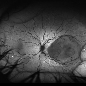
Autofluorescence OD. 35 year-old female with bilateral serous retinal detachment with acute loss of visual acuity.
Photographer: Nicolas Crim MD, Córdoba, Argentina
Condition/keywords: Vogt-Koyanagi-Harada
-
 Serous Retinal Detachment in a patient with Vogt-Koyanagi-Harada syndrome
Aug 24 2021 by Nicolás Crim, MD
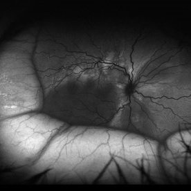
Fundus OD. 35 year-old female with bilateral serous retinal detachment with acute loss of visual acuity
Photographer: Nicolas Crim MD, Córdoba, Argentina
Condition/keywords: serous retinal detachment, vision loss, Vogt-Koyanagi-Harada
-
 Serous Retinal Detachment in a patient with Vogt-Koyanagi-Harada syndrome
Aug 24 2021 by Nicolás Crim, MD
Autofluorescence OS. 35 year-old female with bilateral serous retinal detachment with acute loss of visual acuity.
Photographer: Nicolas Crim MD, Córdoba, Argentina
Condition/keywords: serous retinal detachment, vision loss, Vogt-Koyanagi-Harada
-
 Serous Retinal Detachment in a patient with Vogt-Koyanagi-Harada Syndrome
Aug 24 2021 by Nicolás Crim, MD
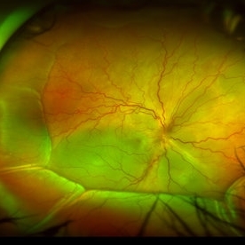
Fundus OS. Female 35 year-old with bilateral Serous Retinal Detachment with acute lost of visual acuity.
Photographer: Nicolas Crim MD, Córdoba, Argentina
Condition/keywords: Vogt-Koyanagi-Harada
-
 Serous Retinal Detachment in Vogt Koyanagi Harada Patient
Apr 26 2021 by Pablo Baquero Ospina, MD
24-year-old woman with bilateral panuveitis and serous retinal detachment, headache and tinnitus.
Photographer: Pablo Baquero-Ospina, Asociación Para Evitar la Ceguera en México
Imaging device: Heidelberg Spectralis
Condition/keywords: serous retinal detachment, Vogt-Koyanagi-Harada
-
 Slide 3-30
Feb 20 2019 by Lancaster Course in Ophthalmology
Low-power view of eye with Vogt-Koyanagi-Harada disease. Note the uveal infiltration.
Condition/keywords: uveal infiltration, Vogt-Koyanagi-Harada
-
 Slide 3-31
Feb 20 2019 by Lancaster Course in Ophthalmology
Higher-power view of choroid in Vogt-Koyanagi-Harada disease ( x65). Note the infiltration of the choroid. In contrast to sympathetic ophthalmia, there is no sparing of the choriocapillaris, and there is destruction of the retinal pigment epithelium.
Condition/keywords: choriocapillaris, choroid, ophthalmia, retinal pigment epithelium, Vogt-Koyanagi-Harada
-
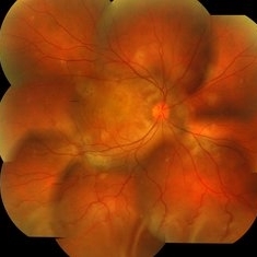
 Sunset Glow Fundus
May 15 2022 by Manuel Ángel Alcántara Delgado, MD
Optomap ultra-widefield retinal imaging of an 35-year-old woman showed sunset glow fundus, multiple nummular chorioretinal atrophic lesions, macular subretinal fibrosis and pigment clumping in chronic recurrent stage of Vogt-Koyanagi-Harada disease.
Photographer: Manuel Ángel Alcántara Delgado. Conde de Valenciana.
Condition/keywords: abnormal retina, benign pigmented lesions, pigment clumps, retinal fibrosis, uveitis, Vogt-Koyanagi-Harada
-
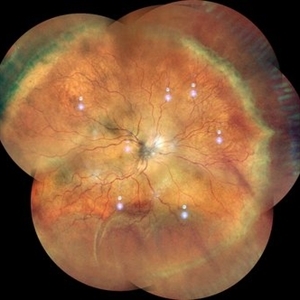 VKH
VKH
Sep 29 2023 by Anjana Mirajkar, MS Ophthalmology
Wide field color photo image of RE of a 41 year old female case of VKH showing exudative retinal detachment inferiorly with multiple fluid pockets in the posterior pole with ILM folds
Photographer: Dr. Anjana Mirajkar -Retina Foundation, Ahmedabad
Imaging device: Mirante-Nidek
Condition/keywords: Vogt-Koyanagi-Harada
-
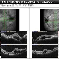 VKH
VKH
Sep 29 2023 by Anjana Mirajkar, MS Ophthalmology
OCT of BE of a 41 year old female in a case of BE VKH showing subretinal fluid with septations with choroidal undulations.
Photographer: Dr. Anjana Mirajkar -Retina Foundation, Ahmedabad
Imaging device: Mirante-Nidek
Condition/keywords: vkh, Vogt-Koyanagi-Harada
-
 VKH
VKH
Sep 29 2023 by Anjana Mirajkar, MS Ophthalmology
Late frame of FA+ICG of RE of a 41 year old female showing disc leakage with hyperfluorescence suggestive of leakage with hypofluoroscence on FA and ICG in a case of VKH.
Photographer: Dr. Anjana Mirajkar -Retina Foundation, Ahmedabad.
Imaging device: Heidelberg
Condition/keywords: Vogt-Koyanagi-Harada
 Loading…