Search results (212 results)
-
 Vogt-Koyanagi-Harada (VKH) Syndrome
Aug 18 2025 by Ricardo Leitão Guerra
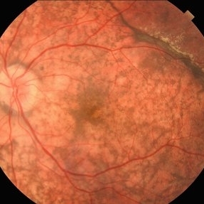
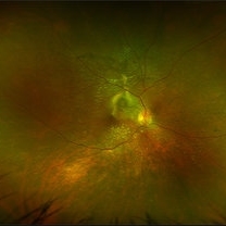
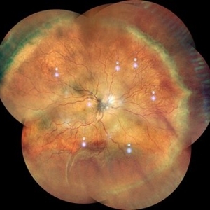
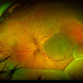
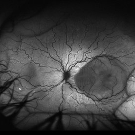
A 45 year old female presenting serous retinal detachment due to Vogt-Koyanagi-Harada (VKH) Syndrome
Photographer: Ricardo Leitão Guerra, Leitão Guerra - Oftalmologia
Imaging device: Zeiss Clarus 700
Condition/keywords: Vogt-Koyanagi-Harada (VKH) Symdrome
-
 Scimitar Sign & Sunset Glow Fundus
Jul 7 2025 by César Adrián Gómez Valdivia, MD
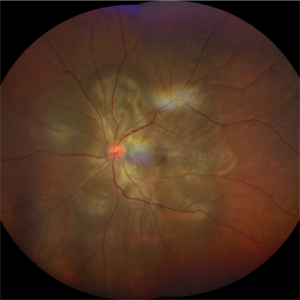
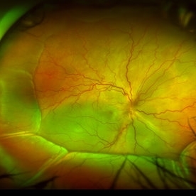
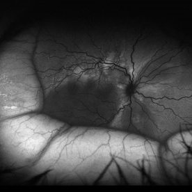
Scimitar Sign & Sunset Glow Fundus found in a 38YO female patient diagnosed with Vogt-Koyanagi-Harada disease. Findings were bilateral.
Photographer: @eyemissu2
Imaging device: TOPCON TRC-50DX
Condition/keywords: Scimitar Sign
-
 VKH Syndrome
Jun 12 2025 by Virginia Gebhart
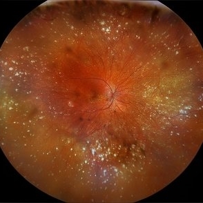
22 year old male with VKH Syndrome. Pt has been experiencing severe headaches, distorted vision, hearing loss, weakness, and a large white patch of hair. Significant cell in AC and vitreous, multiple punched-out CR scars in periphery. Referred to rheumatology for possible immunomodulatory treatment
Photographer: Virginia Gebhart, Retina Consultants of Carolina
Imaging device: Optos California
Condition/keywords: montage, multifocal choroiditis, panuveitis, Vogt-Koyanagi-Harada
-
 VKH Syndrome
Jun 12 2025 by Virginia Gebhart
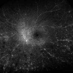
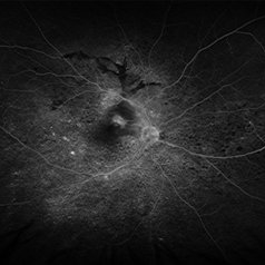
Fluorescein angiogram of 22 year old male with VKH syndrome. Significant cell in AC and vitreous, multiple punched-out CR scars in periphery, mild vascular leakage. Pt referred to rheumatology for immunomodulatory treatment.
Photographer: Virginia Gebhart, Retina Consultants of Carolina
Imaging device: Optos California
Condition/keywords: FA, fluorescein angiogram (FA), multifocal choroiditis, panuveitis, VKH, Vogt-Koyanagi-Harada
-
 Vogt Kayanagi Harada Disease
May 26 2025 by Malvika Singh
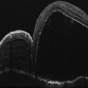
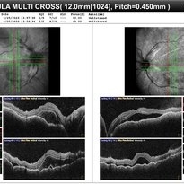
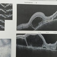
OCT image of the retina showing SRF in case of an exudative retinal detachment and bacillary layer detachment
Photographer: Dr Malvika Singh, Retina Foundation, Ahmedabad, India
Imaging device: Mirante SLO/OCT
Condition/keywords: OCT, Vogt-Koyanagi-Harada
-
 VKH Pseudotumor – Chronic Subretinal Fibrosis
May 11 2025 by Felipe Murati
Ultra-widefield fundus image from a 36-year-old woman with chronic VKH syndrome showing a pseudotumor-like subretinal fibrotic lesion in the right eye. The lesion developed after multiple relapses and remained stable over a 1-year follow-up with immunosuppressive treatment including prednisone, mycophenolate mofetil, and adalimumab. No active choroiditis or exudative detachment was observed. Multimodal imaging was essential for disease monitoring.
Photographer: Felipe A. Murati, MD, University of Arizona
Imaging device: Optos California ultra-widefield retinal imaging system, single-capture, color fundus modality.
Condition/keywords: adalimumab, chronic inflammation, granulomatous uveitis, OCT, Optos ultra-widefield imaging, pseudotumor, subretinal fibrosis, VKH, Vogt-Koyanagi-Harada
-
 VKH Pseudotumor – Fluorescein Angiography
May 11 2025 by Felipe Murati
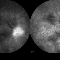
Fluorescein angiography image from a 36-year-old woman with chronic Vogt-Koyanagi-Harada (VKH) syndrome showing a pseudotumor-like lesion with late-phase staining and no active leakage. The image highlights subretinal fibrosis in the right eye, stable under long-term immunosuppressive therapy with mycophenolate mofetil and adalimumab. No signs of active choroiditis are present, confirming a quiescent phase.
Photographer: Felipe A. Murati, MD, University of Arizona
Imaging device: Optos California, fluorescein angiography modality
Condition/keywords: choroiditis, Fluorescein angiography, granulomatous uveitis, Optos FA, pseudotumor, subretinal fibrosis, VKH, Vogt-Koyanagi-Harada
-
 Serous Retinal Detachment
Oct 14 2024 by César Adrián Gómez Valdivia, MD
Serous Retinal Detachment found in a 19 year-old female patient with suspected Vogt-Koyanagi-Harada disease. Findings were bilateral. Patient was admitted for Methylprednisolone and Cyclophosphamide treatment.
Photographer: @eyemissu2
Imaging device: California ICG OPTOS
Condition/keywords: serous retinal detachment, vkh, Vogt-Koyanagi-Harada
-
 Serous Retinal Detachment
Oct 14 2024 by César Adrián Gómez Valdivia, MD
Serous Retinal Detachment found in a 19 year-old female patient with suspected Vogt-Koyanagi-Harada disease. Findings were bilateral. Patient was admitted for Methylprednisolone and Cyclophosphamide treatment.
Photographer: @eyemissu2
Imaging device: California ICG OPTOS
Condition/keywords: serous retinal detachment, Vogt-Koyanagi-Harada
-
 Vogt-Koyanagi-Harada Disease
Sep 24 2024 by Gustavo Uriel Fonseca Aguirre
A 39-year-old female patient with no ophthalmologic history was diagnosed with Vogt-Koyanagi-Harada disease. Exudative retinal detachment was observed in the left eye.
Photographer: Gustavo U. Fonseca Aguirre, Fundación Hospital Nuestra Señora de la Luz, Ciudad de México
Condition/keywords: Vogt-Koyanagi-Harada
-
 Vogt-Koyanagi-Harada Disease
Sep 24 2024 by Gustavo Uriel Fonseca Aguirre
A 39-year-old female patient with no ophthalmologic history was diagnosed with Vogt-Koyanagi-Harada disease. Exudative retinal detachment was observed in the right eye.
Photographer: Gustavo U. Fonseca Aguirre, Fundación Hospital Nuestra Señora de la Luz, Ciudad de México
Condition/keywords: Vogt-Koyanagi-Harada
-
 VKH
VKH
Sep 29 2023 by Anjana Mirajkar, MS Ophthalmology
Late frame of FA+ICG of RE of a 41 year old female showing disc leakage with hyperfluorescence suggestive of leakage with hypofluoroscence on FA and ICG in a case of VKH.
Photographer: Dr. Anjana Mirajkar -Retina Foundation, Ahmedabad.
Imaging device: Heidelberg
Condition/keywords: Vogt-Koyanagi-Harada
-
 VKH
VKH
Sep 29 2023 by Anjana Mirajkar, MS Ophthalmology
OCT of BE of a 41 year old female in a case of BE VKH showing subretinal fluid with septations with choroidal undulations.
Photographer: Dr. Anjana Mirajkar -Retina Foundation, Ahmedabad
Imaging device: Mirante-Nidek
Condition/keywords: vkh, Vogt-Koyanagi-Harada
-
 VKH
VKH
Sep 29 2023 by Anjana Mirajkar, MS Ophthalmology
Wide field color photo image of RE of a 41 year old female case of VKH showing exudative retinal detachment inferiorly with multiple fluid pockets in the posterior pole with ILM folds
Photographer: Dr. Anjana Mirajkar -Retina Foundation, Ahmedabad
Imaging device: Mirante-Nidek
Condition/keywords: Vogt-Koyanagi-Harada
-
 VKH - Uveitic stage
Jun 23 2023 by Sergio Emilio Sifuentes Renteria, MD
Fundus photograph of a young female with VKH in uveitic stage
Photographer: Sergio Emilio Sifuentes Rentería - Foundation Hospital Nuestra Señora de La Luz
Condition/keywords: choroiditis, panuveitis, serous retinal detachment, VKH, Vogt-Koyanagi-Harada
-
 Sunset Glow Fundus
May 15 2022 by Manuel Ángel Alcántara Delgado, MD
Optomap ultra-widefield retinal imaging of an 35-year-old woman showed sunset glow fundus, multiple nummular chorioretinal atrophic lesions, macular subretinal fibrosis and pigment clumping in chronic recurrent stage of Vogt-Koyanagi-Harada disease.
Photographer: Manuel Ángel Alcántara Delgado. Conde de Valenciana.
Condition/keywords: abnormal retina, benign pigmented lesions, pigment clumps, retinal fibrosis, uveitis, Vogt-Koyanagi-Harada
-
 Vogt-Koyanagi-Harada Disease
Apr 24 2022 by Aniruddha K Agarwal, MD
A 38-year-old woman of Asian descent with no ophthalmological or systemic history presented to the emergency eye clinic with a 1-week complaint of headache and bilateral vision loss. Funduscopy revealed bilateral serous neurosensory detachments. The presence of lymphocytosis in cerebrospinal fluid and mild acute sensorineural hearing loss confirmed the diagnosis of uveomeningoencephalitic syndrome (Vogt-Koyanagi-Harada disease).
Photographer: Mercedes SERRADOR, MD, PhD and Beatriz VENTAS, MD
Imaging device: Zeiss Clarus fundus camera
Condition/keywords: IUSG, panuveitis, Vogt-Koyanagi-Harada
-
 Recurrent VKH
Mar 8 2022 by mohamed ahmed el shafie, ophthalmic resident
A case of recurrent VKH. OCT scan
Condition/keywords: subretinal fluid, Vogt-Koyanagi-Harada
-
 Stage 3 Vogt-Koyanagi-Harada (VKH) Disease
Oct 20 2021 by Bryon R McKay, MD, PhD, FRCSC, DRCPSC - Retina
27yF presented with sub-acute findings of VKH, she has an interesting pattern of perivascular changes. She was successfully treated with immunosuppressive agents and maintains 20/20 vision.
Photographer: Dr. Vaezi, University of British Columbia
Imaging device: Optos Imaging System
Condition/keywords: uveitis
-
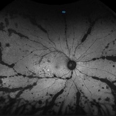 Autofluorescence Stage 3 Vogt-Koyanagi-Harada (VKH) Disease
Oct 20 2021 by Bryon R McKay, MD, PhD, FRCSC, DRCPSC - Retina
27yF presented with sub-acute findings of VKH, she has an interesting pattern of perivascular changes. She was successfully treated with immunosuppressive agents and maintains 20/20 vision.
Photographer: Dr. K. Vaezi, University of British Columbia, Canada
Imaging device: Optos Imaging system
Condition/keywords: Vogt-Koyanagi-Harada
-
 2min-FA-VKH
Oct 20 2021 by Bryon R McKay, MD, PhD, FRCSC, DRCPSC - Retina
27yF presented with sub-acute findings of VKH, she has an interesting pattern of perivascular changes. She was successfully treated with immunosuppressive agents and maintains 20/20 vision.
Photographer: Dr. K. Vaezi, University of British Columbia, Canada
Imaging device: Optos Imaging system
Condition/keywords: uveitis, Vogt-Koyanagi-Harada
-
 Serous Retinal Detachment in a patient with Vogt-Koyanagi-Harada Syndrome
Aug 24 2021 by Nicolás Crim, MD
Fundus OS. Female 35 year-old with bilateral Serous Retinal Detachment with acute lost of visual acuity.
Photographer: Nicolas Crim MD, Córdoba, Argentina
Condition/keywords: Vogt-Koyanagi-Harada
-
 Serous Retinal Detachment in a patient with Vogt-Koyanagi-Harada syndrome
Aug 24 2021 by Nicolás Crim, MD
Autofluorescence OS. 35 year-old female with bilateral serous retinal detachment with acute loss of visual acuity.
Photographer: Nicolas Crim MD, Córdoba, Argentina
Condition/keywords: serous retinal detachment, vision loss, Vogt-Koyanagi-Harada
-
 Serous Retinal Detachment in a patient with Vogt-Koyanagi-Harada syndrome
Aug 24 2021 by Nicolás Crim, MD
Fundus OD. 35 year-old female with bilateral serous retinal detachment with acute loss of visual acuity
Photographer: Nicolas Crim MD, Córdoba, Argentina
Condition/keywords: serous retinal detachment, vision loss, Vogt-Koyanagi-Harada
-
 Serous Retinal Detachment in a patient with Vogt-Koyanagi-Harada Syndrome
Aug 24 2021 by Nicolás Crim, MD
Autofluorescence OD. 35 year-old female with bilateral serous retinal detachment with acute loss of visual acuity.
Photographer: Nicolas Crim MD, Córdoba, Argentina
Condition/keywords: Vogt-Koyanagi-Harada
 Loading…