Search results (155 results)
-
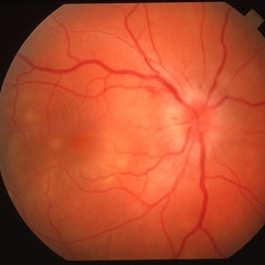
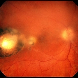 Toxoplasma Retinochoroiditis
Feb 25 2013 by Henry J. Kaplan, MD
Toxoplasmosis, right eye: reactivation of congenital toxoplasmosis as an active retinitis lesion with overlying vitritis adjacent to an old scar.
Condition/keywords: toxoplasmosis chorioretinitis, toxoplasmosis reactivation
-
 Candida Vitritis
Oct 18 2012 by Larry Halperin, MD
Candida vitritis
Condition/keywords: candida endophthalmitis, vitritis
-
---thumb.jpg/image-square;max$300,300.ImageHandler) Multifocal Choroiditis and Panuveitis Syndrome
Multifocal Choroiditis and Panuveitis Syndrome
Feb 26 2013 by Henry J. Kaplan, MD
Multifocal choroiditis and panuveitis: left eye. Acute stage: haziness of the media due to vitritis and multiple active yellow and also inactive choroidal lesions are present.
Condition/keywords: multifocal choroiditis
-
---thumb.jpg/image-square;max$300,300.ImageHandler) Multifocal Choroiditis & Panuveitis Syndrome
Multifocal Choroiditis & Panuveitis Syndrome
Feb 26 2013 by Henry J. Kaplan, MD
Multifocal choroiditis, MFC. Lesions look similar to POHS but the patient has vitritis in contrast to the former.
Condition/keywords: multifocal choroiditis, panuveitis
-
---thumb.jpg/image-square;max$300,300.ImageHandler) Ocular Toxoplasmosis
Ocular Toxoplasmosis
Feb 15 2013 by From the Collections of Thomas M. Aaberg, MD and Thomas M. Aaberg Jr., MD
Diffuse slit-lamp photograph of the right eye of a patient with ocular toxoplasmosis showing strands of vitreous inflammation.
Condition/keywords: ocular toxoplasmosis, vitritis
-
 Toxoplasma Gondii Chorioretinitis
Aug 23 2012 by Gerardo Garcia-Aguirre, MD
Fundus photograph of the superior periphery showing vitritis, and an atrophic lesion with an adjacent focus of chorioretinitis.
Photographer: Noemí Hernández, Asociación para Evitar la Ceguera en México
Imaging device: Zeiss FF4
Condition/keywords: toxoplasmosis
-
 Syphilitic Panuveitis, Left Eye
Oct 8 2012 by Pauline T Merrill, MD, FASRS
Left fundus photograph of a 23-year-old Hispanic male with decreased vision for 1 month, recent pain/redness both eyes. Found to have panuveitis; also rash on palms & soles. Labs positive for FTA-ABS, and CSR VDRL. Treated with IV penicillin for 14 days.
Photographer: Karen Parque, Illinois Retina Associates, Chicago, IL
Condition/keywords: panuveitis, snowballs, syphilis, vitritis
-
 Acute Retinal Necrosis (ARN)
Dec 13 2017 by Gabriel Costa Andrade, PhD
Healthy 47-year-old patient presenting with subacute decline in vision, vitritis, periphlebits, and necrotizing retinitis.
Photographer: Gabriel Andrade, RETINA CLINIC, SP
Imaging device: Optos Wide Field Camera
Condition/keywords: acute retinal necrosis, uveitis, vasculitis
-
 Syphilitic Panuveitis, Right Eye
Oct 8 2012 by Pauline T Merrill, MD, FASRS
Right fundus photograph of a 23-year-old Hispanic male with decreased vision for 1 month, recent pain/redness both eyes. Found to have panuveitis; also rash on palms & soles. Labs positive for FTA-ABS, and CSR VDRL. Treated with IV penicillin for 14 days.
Photographer: Karen Parque, Illinois Retina Associates, Chicago, IL
Condition/keywords: panuveitis, snowballs, syphilis, vitritis
-
 Toxoplasmosis Slide 1
Oct 22 2012 by Ronald C. Gentile, MD
35-year-old women presented with decreasing vision in the left eye with progressive central scotoma. Fundus examination revealed one focal area of chorioretinitis adjacent to one of multiple old pigmented retinal scars. The focal area of chorioretinitis involved the deep retinal layers and was associated with sub-retinal fluid and little overlying vitritis.
Photographer: The New York Eye & Ear Infirmary Department of Medical Imaging
Condition/keywords: punctate outer retinal toxoplasmosis, toxoplasmosis
-
---thumb.jpg/image-square;max$300,300.ImageHandler) ARN
ARN
Feb 27 2013 by Henry J. Kaplan, MD
ARN, severe vitritis, and necrotizing retina.
Condition/keywords: acute retinal necrosis, necrotizing retina, vitritis
-
 Asteroid Hyalosis, Vitreous Face Attached
Dec 10 2012 by Yale L. Fisher, MD
In asteroid hyalosis, accumulations of calcium soaps dispersed throughout the vitreous produce bright echoes in the usually echolucent vitreous. The appearance of asteroid hyalosis should not be confused with that of vitreous hemorrhage or vitritis. Many of the larger aggregates in asteroid hyalosis are easily seen as the gain is reduced to below 60 db, unlike vitreous hemorrhage or vitritis which usually disappears at low gain settings. There is also an area of clear echolucent vitreous between the posterior hyaloid face and the asteroid particles, which is usually not present in vitreous hemorrhage or vitritis.
Condition/keywords: video
-
---thumb.jpg/image-square;max$300,300.ImageHandler) Birdshot Retinochoroidopathy
Birdshot Retinochoroidopathy
Feb 26 2013 by Henry J. Kaplan, MD
Birdshot retinochoroidopathy; left eye: acute stage, haze due to vitritis and multiple oval cream colored lesions at the level of choroid.
Condition/keywords: birdshot retinochoroidopathy
-
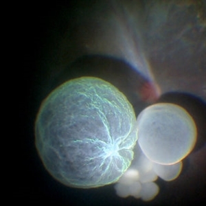 Intraocular Multiple Cysticercus
Oct 10 2018 by Vishal Agrawal, MD, FRCS,FACS,FASRS
Intraoperative fundus picture of right eye of a 18-year-old boy with complaints of DOV for the past 2 months. There were 12 intravitreal cysts in total with vitritis sclerosis retinal vessels and TRD. To note here, the largest cyst has a flimsy wall and no scolex (possibly ruptured) and the rest of the smaller cysts have a scolex and a taut wall.
Photographer: Vishal Agrawal MD,FRCS
Imaging device: SONY PMW-10 MD HD
Condition/keywords: cysticercosis, scolex
-
 vitreous exudate
Feb 15 2013 by From the Collections of Thomas M. Aaberg, MD and Thomas M. Aaberg Jr., MD
Gross pathologic specimen showing vitreous organization and opacities consistent with severe intraocular inflammation.
Condition/keywords: gross pathology, vitritis
-
---thumb.jpg/image-square;max$300,300.ImageHandler) ARN
ARN
Feb 27 2013 by Henry J. Kaplan, MD
ARN; hazy media is due to moderate to severe vitritis
Condition/keywords: acute retinal necrosis
-
---thumb.jpg/image-square;max$300,300.ImageHandler) Lyme Disease
Lyme Disease
Feb 26 2013 by Henry J. Kaplan, MD
Lyme disease, intermediate uveitis: severe vitritis and snow ball formations. #6
Condition/keywords: Lyme disease
-
---thumb.jpg/image-square;max$300,300.ImageHandler) vitreous haze and confluent peripheral retinal whitening consistent with active ocular toxoplasmosis
vitreous haze and confluent peripheral retinal whitening consistent with active ocular toxoplasmosis
Feb 15 2013 by From the Collections of Thomas M. Aaberg, MD and Thomas M. Aaberg Jr., MD
Color fundus photograph showing vitreous haze and confluent peripheral retinal whitening consistent with active ocular toxoplasmosis
Condition/keywords: ocular toxoplasmosis, vitritis
-
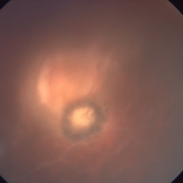
 'Headlight in the Fog' in Toxoplasmosis
Apr 8 2019 by Gary R. Cook, MD, FACS
29-year-old white male with moderate vitritis associated with an active toxoplasmosis lesion OS resulting in a "headlight in the fog" picture.
Condition/keywords: toxo chorioretinitis, toxoplasmosis, vitritis
-
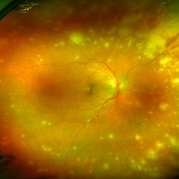
 Posterior Uveitis
Apr 8 2019 by Gary R. Cook, MD, FACS
37-year-old white male with mild vitritis, optic disc hyperemia and edema, peripapillary hemorrhages and yellow-white spots in temporal macula OD; V.A. = 20/30.
Imaging device: Topcon VT-50
Condition/keywords: posterior uveitis
-
---thumb.jpg/image-square;max$300,300.ImageHandler) Vitreous haze and confluent peripheral retinal whitening
Vitreous haze and confluent peripheral retinal whitening
Feb 15 2013 by From the Collections of Thomas M. Aaberg, MD and Thomas M. Aaberg Jr., MD
Color fundus photograph showing vitreous haze and confluent peripheral retinal whitening consistent with active ocular toxoplasmosis.
Condition/keywords: ocular toxoplasmosis, vitritis
-
 Toxoplasmosis Retinochoroiditis
Feb 24 2014 by Susanna S. Park, MD, PhD
Fundus photo of a 35-year-old woman with new vision loss and floaters showing focal retinochoroiditis adjacent to an old scar and vitritis.
Photographer: Ellen Redenbo, University of California Davis
Condition/keywords: acute, toxoplasmosis chorioretinitis, vitritis
-
 Toxoplasmosis
Feb 24 2014 by Susanna S. Park, MD, PhD
Posterior pole fundus photo of a 35-year-old woman showing media haze from acute vitritis associated with a focal mid-peripheral retinochorioretinitis next to an old scar.
Photographer: Ellen Redeenbo, University of California Davis Eye Center
Condition/keywords: toxoplasmosis chorioretinitis, vitritis
-
---thumb.jpg/image-square;max$300,300.ImageHandler) Vitreous haze and focal peripheral retinal whitening consistent with active ocular toxoplasmosis
Vitreous haze and focal peripheral retinal whitening consistent with active ocular toxoplasmosis
Feb 15 2013 by From the Collections of Thomas M. Aaberg, MD and Thomas M. Aaberg Jr., MD
Color fundus photograph showing vitreous haze and focal peripheral retinal whitening consistent with active ocular toxoplasmosis
Condition/keywords: ocular toxoplasmosis, vitritis
-
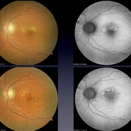 Aquired Vitelliform Maculopathy
Jun 29 2014 by John S. King, MD
Asymptomatic MA healthy female consulted for wet AMD. Plan: observe. Photo and AF initially and year afterwards. Health MA female; no vitritis or other lesions; similar findings in both eyes.
Photographer: Wayne A Ladlee Jr
Condition/keywords: aquired vitelliform maculopathy
 Loading…