Search results (39 results)
-
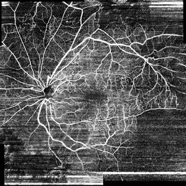
 Whole Eye OCT
Jan 4 2019 by Netan Choudhry, MD, FRCS(C) FASRS
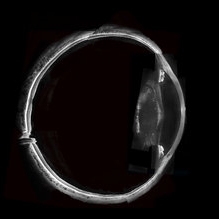
Swept-Source OCT montage of a 45-year-old male with Alports disease and posterior subcapsular cataract.
Photographer: John Golding BA, Vitreous Retina Macula Specialists of Toronto
Imaging device: Topcon DRI Triton
Condition/keywords: Alports disease, optical coherence tomography (OCT), swept source
-
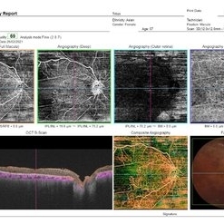
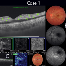 Figure-1 Paracentral Acute Middle Maculopathy (PAMM)
Dec 21 2018 by Fawwaz F Al Mamoori, MD, Medical Retina Consultant
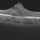
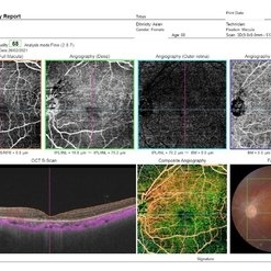
25-year-old male patient medically free, had sudden deterioration in his left eye vision. Visual acuity on presentation was counting fingers at 3 meter distance. Marked Relative Pupillary Afferent Defect (RAPD) was detected and fundoscopic exam showed abnormal foveal reflex. SS OCT B scan: showed a hypereflectivity of the inner plexiform layer (IPL), inner nuclear layer (INL) and OPL layer (fig 1, A).FA images were normal (fig 1, B). Angiography shows remarkable perifoveal capillary drop out within middle retinal layer correlating with perfusion density map which reveals significant decrease in capillary density at the same level (Fig 1, C). Enface ads more proof to PAMM by delineating ischemic distribution in a fern like pattern of hyper reflective areas within DCP (fig1, D).
Photographer: Dr.Fawwaz Al Mamoori (Al Mamoori Eye Clinic)
Imaging device: Triton Swept Source OCT (TOPCON)
Condition/keywords: optical coherence tomography (OCT), paracentral acute middle maculopathy
-
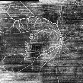
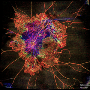 Flame of the Forest
Apr 9 2020 by Daraius N Shroff, MS FMRF FRCS
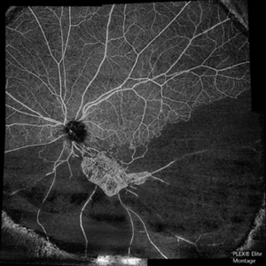
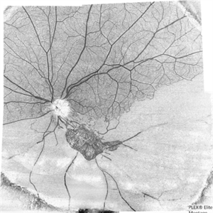
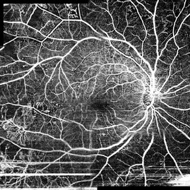
A 54-year-old man with DM for 15 years. The left eye had a visual acuity of 20/40. Wide field swept source OCTA revealed branching out central neovascular trunk vessels from the disc with terminal loops, along with exuberant proliferation of irregular small-calibre fine new vessels. The patient underwent OCTA guided pan retinal photocoagulation.
Photographer: Anuj Choudhary, Shroff Eye Centre, New Delhi
Imaging device: Zeiss Plex Elite 9000
Condition/keywords: proliferative diabetic retinopathy (PDR)
-
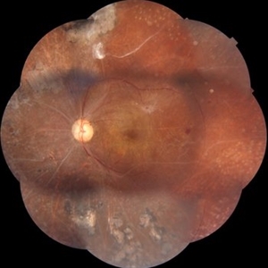
 Optic Disc Pit Maculopathy
Aug 20 2018 by DIEGO A BUESO PONCE, MD
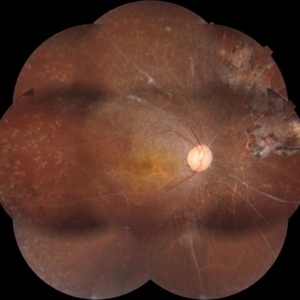
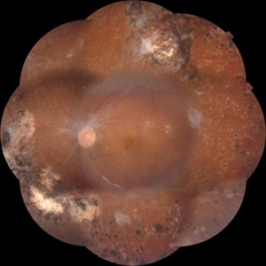
Fundus photograph of an 19-year-old female with congenital optic disc pit and associated maculopathy with subretinal fluid and retinoschisis.
Photographer: Diego Bueso Ponce, Clinica Unidad Laser, Barranquilla Colombia
Imaging device: Topcon DRI OCT Triton, Swept source OCT
Condition/keywords: congenital optic nerve pit, maculopathy, optic disc pit, retinoschisis
-
 Figure-2 Paracentral Acute Middle Maculopathy (PAMM)
Dec 21 2018 by Fawwaz F Al Mamoori, MD, Medical Retina Consultant
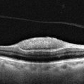
70-year-old female patient known to have hypertension, presented with acute deterioration of left eye vision, best corrected visual acuity was 6/60. Fundoscopic exam showed abnormal foveal reflex whiting .SS-OCT B scan showed also a hypereflectivity of the inner plexiform layer (IPL), inner nuclear layer (INL) and OPL layer(figure-2, A). FA images were also normal(figure-2 B). Segmented angiographic images elucidate ischemia and capillary drop out predominantly at the level of DCP but less severe than Case 1 (fig 2, C). Correspondingly, Enface highlights hyper reflective areas in a fern like distribution in the middle retina at similar depth of ischemic lesions demonstrated on B scans and OCTA (fig 2, D)
Photographer: Dr.Fawwaz Al Mamoori (Al Mamoori Eye Clinic)
Imaging device: Triton Swept Source OCT (TOPCON)
Condition/keywords: optical coherence tomography (OCT), paracentral acute middle maculopathy
-
 Astrocytoma OCT
Jan 9 2018 by Sidra Zafar
Swept source OCT imaging of retinal astrocytoma in a female child with known diagnosis of tuberous sclerosis.
Imaging device: Swept Source
Condition/keywords: astrocytoma, optical coherence tomography (OCT), tuberous sclerosis
-
 Astrocytoma OCT
Jan 9 2018 by Sidra Zafar
OCT imaging of retinal astrocytoma in a female child with known diagnosis of tuberous sclerosis. A cystic pocket can be observed.
Imaging device: Swept Source
Condition/keywords: tuberous sclerosis
-
 OCT Choroidal Tuberculoma
Jun 15 2020 by Aayesha Khanum
OCT choroidal tuberculoma.
Photographer: Puttaswamy
Imaging device: DRI OCT Triton swept source OCT-Topcon
Condition/keywords: abscess, choroidal tuberculoma
-
 Choroidal Tuberculoma
Jun 15 2020 by Aayesha Khanum
Fundus photo of diagnosed case of pulmonary TB.
Photographer: Puttaswamy
Imaging device: DRI OCT Triton swept source OCT- Topcon
Condition/keywords: abscess, choroidal tuberculoma
-
 The Barren Field
Jun 26 2020 by SANDEEP KUMAR
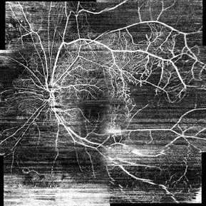
A 59-year-old man with DM for 18 years operated for mature cataract. Post op left eye had a visual acuity of 20/80. Wide field swept source OCTA revealed gross vessel wipe out in inferior hemi quadrant with branching out neovascular frond inferior to disc with terminal loops, The patient underwent Anti VEGF injection followed by OCTA guided sectoral retinal photocoagulation.Image J software used here to generate reverse image that sharply delineates the non perfusion are
Photographer: Sandeep Kumar
Imaging device: Optical coherence tomography system Zeiss Plex Elite 9000
Condition/keywords: hemi CRVO
-
 OCT of a sub-internal limiting membrane hemorrhage in Valsalva retinopathy
Jul 29 2022 by JORGE SOBERANES
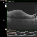
Optical coherence tomography of a 70-year-old man with a sub-internal limiting membrane due to Valsalva retinopathy
Photographer: Jorge I. Soberanes, Asociación para Evitar la Ceguera en México.
Imaging device: PLEX Elite 9000, Zeiss
Condition/keywords: OCT, sub-inner limiting membrane hemorrhage, swept source, valsalva retinopathy
-
 Proliferative Diabetic Retinopathy
Mar 1 2021 by Avris Romario Diparaja Siahaan
Fundus photograph (montage photography) of a 57-year-old woman with proliferative diabetic retinopathy in her both eyes.
Photographer: Nanda Lessi Hafni Eka Putri, MD (Ophthalmologist) & Ryan Mishbahuddin (Nurse), Ciawi General Hospital (Rumah Sakit Umum Daerah Ciawi)
Imaging device: DRI OCT Triton Plus
Condition/keywords: fundus photograph, montage, optical coherence tomography (OCT), swept source, wide angle imaging
-
 Proliferative Diabetic Retinopathy
Mar 1 2021 by Avris Romario Diparaja Siahaan
Swept source OCT angiography (12.0 mm X 12.0 mm) of a 57-year-old woman with proliferative diabetic retinopathy in her both eyes.
Photographer: Nanda Lessi Hafni Eka Putri, MD (Ophthalmologist) & Ryan Mishbahuddin (Nurse), Ciawi General Hospital (Rumah Sakit Umum Daerah Ciawi)
Imaging device: DRI OCT Triton Plus
Condition/keywords: fundus photograph, montage, optical coherence tomography (OCT), swept source, wide angle imaging
-
 Proliferative Diabetic Retinopathy
Mar 1 2021 by Avris Romario Diparaja Siahaan
Fundus photograph (montage photography) of a 57-year-old woman with proliferative diabetic retinopathy in her both eyes.
Photographer: Nanda Lessi Hafni Eka Putri, MD (Ophthalmologist) & Ryan Mishbahuddin (Nurse), Ciawi General Hospital (Rumah Sakit Umum Daerah Ciawi)
Imaging device: DRI OCT Triton Plus
Condition/keywords: fundus photograph, montage, optical coherence tomography (OCT), swept source, wide angle imaging
-
 Proliferative Diabetic Retinopathy
Mar 1 2021 by Avris Romario Diparaja Siahaan
Swept Source OCT angiography (montage photography) of a 57-year-old woman with proliferative diabetic retinopathy in her both eyes.
Photographer: Nanda Lessi Hafni Eka Putri, MD (Ophthalmologist) & Ryan Mishbahuddin (Nurse), Ciawi General Hospital (Rumah Sakit Umum Daerah Ciawi)
Imaging device: DRI OCT Triton Plus
Condition/keywords: fundus photograph, montage, optical coherence tomography (OCT), swept source, wide angle imaging
-
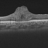
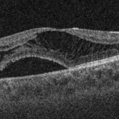 Retinoschisis and Subretinal Fluid in Optic Disc Pit Related Maculopathy
Aug 20 2018 by DIEGO A BUESO PONCE, MD
OCT B-scan of a 19-year-old female with congenital optic disc pit maculopathy.
Photographer: Diego Bueso Ponce, Clinica Unidad Laser, Barranquilla Colombia
Imaging device: Topcon DRI OCT Triton, Swept source OCT
Condition/keywords: B scan ultrasound, congenital optic nerve pit, retinoschisis
-
 The Barren Field
Jun 27 2020 by SANDEEP KUMAR
A 59-year-old man with DM for 18 years operated for mature cataract. Post op left eye had a visual acuity of 20/80. Wide field swept source OCTA revealed gross vessel wipe out in inferior hemi quadrant with branching out neovascular frond inferior to disc with terminal loops, The patient underwent Anti VEGF injection followed by OCTA guided sectoral retinal photocoagulation.Image J software used here to generate reverse image that sharply delineates the non perfusion area.
Photographer: Sandeep Kumar
Imaging device: Optical coherence tomography system Zeiss Plex Elite 9000
Condition/keywords: hemi CRVO, neovascularization elsewhere (NVE)
-
 Proliferative Diabetic Retinopathy
Mar 1 2021 by Avris Romario Diparaja Siahaan
Swept source OCT angiography (montage photography) of a 57-year-old woman with proliferative diabetic retinopathy in her both eyes.
Photographer: Nanda Lessi Hafni Eka Putri, MD (Ophthalmologist) & Ryan Mishbahuddin (Nurse), Ciawi General Hospital (Rumah Sakit Umum Daerah Ciawi)
Imaging device: DRI OCT Triton Plus
Condition/keywords: fundus photograph, montage, optical coherence tomography (OCT), swept source, wide angle imaging
-
 Retinal Hemorrhage
Sep 2 2021 by Avris Romario Diparaja Siahaan
Swept source OCT angiography of a 58-year-old man with hemorrhage in his left eye.
Photographer: Nanda Lessi Hafni Eka Putri, MD (Ophthalmologist) & Ryan Mishbahuddin (Nurse), Ciawi General Hospital (Rumah Sakit Umum Daerah Ciawi)
Imaging device: DRI OCT Triton Plus (Topcon)
Condition/keywords: fundus photograph, optical coherence tomography (OCT)
-
 Proliferative Diabetic Retinopathy
Mar 1 2021 by Avris Romario Diparaja Siahaan
Swept Source OCT angiography (9.0 mm X 9.0 mm) of a 62-year-old woman with proliferative diabetic retinopathy in her both eyes.
Photographer: Nanda Lessi Hafni Eka Putri, MD (Ophthalmologist) & Ryan Mishbahuddin (Nurse), Ciawi General Hospital (Rumah Sakit Umum Daerah Ciawi)
Imaging device: DRI OCT Triton Plus
Condition/keywords: fundus photograph, montage, optical coherence tomography (OCT), swept source, wide angle imaging
-
 Proliferative Diabetic Retinopathy
Mar 1 2021 by Avris Romario Diparaja Siahaan
Swept source OCT angiography (montage photography) of a 62-year-old woman with proliferative diabetic retinopathy in her both eyes.
Photographer: Nanda Lessi Hafni Eka Putri, MD (Ophthalmologist) & Ryan Mishbahuddin (Nurse), Ciawi General Hospital (Rumah Sakit Umum Daerah Ciawi)
Imaging device: DRI OCT Triton Plus
Condition/keywords: fundus photograph, montage, optical coherence tomography (OCT), swept source, wide angle imaging
-
 Resolving of Refractory Subretinal Fluid Post Faricimab Injection
Nov 7 2022 by Fawwaz F Al Mamoori, MD, Medical Retina Consultant
A case of Rt eye wet-AMD treated over 2 years with a Q8weeks aflibercebt injection with refractory SRF less than 200 um that shifted recently to Faricimab.1 month post Faricimab OCT showed a complete drying out of refractory subretinal Fluid (SRF)
Photographer: Mohammed Rabaa
Imaging device: Swept Source OCT (TRITON) Topcon
Condition/keywords: subretinal fluid
-
 Proliferative Diabetic Retinopathy
Mar 1 2021 by Avris Romario Diparaja Siahaan
Fundus photograph (montage) of a 62-year-old woman with proliferative diabetic retinopathy in her both eyes.
Photographer: Nanda Lessi Hafni Eka Putri, MD (Ophthalmologist) & Ryan Mishbahuddin (Nurse), Ciawi General Hospital (Rumah Sakit Umum Daerah Ciawi)
Imaging device: DRI OCT Triton Plus
Condition/keywords: fundus photograph, montage, optical coherence tomography (OCT), swept source, wide angle imaging
-
 Lung Cancer Choroidal Metastases
Aug 20 2018 by DIEGO A BUESO PONCE, MD
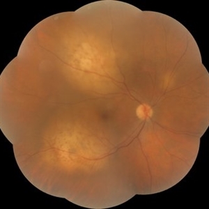
Fundus photograph of an 52-year-old female with multiple choroidal metastases from primary lung cancer.
Photographer: Diego Bueso Ponce, Clinica Unidad Laser, Barranquilla Colombia
Imaging device: Topcon DRI OCT Triton Swept source OCT
Condition/keywords: choroidal metastasis
-
 Proliferative Diabetic Retinopathy
Mar 1 2021 by Avris Romario Diparaja Siahaan
Swept source OCT angiography (montage photography) of a 62-year-old woman with proliferative diabetic retinopathy in her both eyes.
Photographer: Nanda Lessi Hafni Eka Putri, MD (Ophthalmologist) & Ryan Mishbahuddin (Nurse), Ciawi General Hospital (Rumah Sakit Umum Daerah Ciawi)
Imaging device: DRI OCT Triton Plus
Condition/keywords: fundus photograph, montage, optical coherence tomography (OCT), swept source, wide angle imaging
 Loading…