Search results (118 results)
-
 Shafer's Sign
Jan 3 2020 by Manuel Ángel Alcántara Delgado, MD
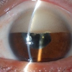
Slit lamp photograph of a 58-year-old man with rhegmatogenous retinal detachment and tobacco dust presence.
Photographer: Manuel Ángel Alcántara Delgado, CMN SXXI, Mexico City
Condition/keywords: acute retinal detachment, retina surgery, vitrectomy
-
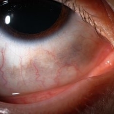 Ocular Melanocytosis Scleral Pigmentation
Jul 9 2014 by Susanna S. Park, MD, PhD
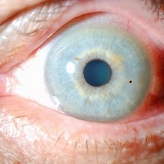
Slit lamp photograph of a 12-year-old boy with ocular melanocytosis showing scleral and episcleral pigmentation. The other eye is blue.
Photographer: Ellen Redenbo
Condition/keywords: melanocytoma
-
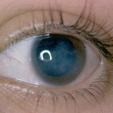
 Ciliary Body Melanoma With Partial Ring Configuration and Diffuse Sentinel Vessels
Feb 26 2014 by Susanna S. Park, MD, PhD
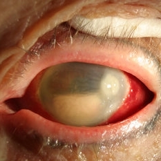
Slit lamp photo of a 57-year-old man with new vision loss from cataract formation. Large ciliary body mass with diffuse sentinel vessels is noted. The eye was removed and the tumor was noted to have a partial ring configuration with predominantly epithelioid cells and early vitreous seeding.
Photographer: Ellen Redenbo, University of California Davis Eye Center
Condition/keywords: ciliary body melanoma, melanoma
-
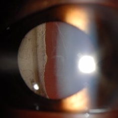
 Glaukomflecken
Oct 23 2017 by Claire Kiernan, MD
Slit lamp photograph of a 59-year-old man with recent-onset severe eye pain noted to have glaukomflecken consistent with recent episode of angle closure glaucoma.
Photographer: Steve Moser, University of Tennessee Hamilton Eye Institute; Joe Mastellone, University of Tennessee Hamilton Eye Institute
Condition/keywords: angle-closure glaucoma interval, glaucoma anterior segment anomalies
-
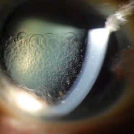 Retention of Perfluorocarbon in Anterior Chamber
Mar 1 2017 by Philip J. Polkinghorne, MD
Slit lamp photograph takien one week after retinal detachment surgery where perfluorocarbon liquid was used to re-attach the retina.
Photographer: Alex Fraser
Condition/keywords: perfluorocarbon fluid, retained perfluorocarbon, retina surgery complications
-
 Niemann Pick Disease Type B
Aug 6 2013 by Hamid Ahmadieh, MD
Photo slit lamp photograph the left eye of a patient with Niemann Pick Type B with corneal stromal depositions.
Photographer: Ali Mohammad-Rabie, Ophthalmic Research Center, Labbafinejad Medical Center, Tehran
Condition/keywords: slit lamp photo
-
---thumb.JPG/image-square;max$300,300.ImageHandler) Carotid Cavernous Fistula
Carotid Cavernous Fistula
Jul 29 2013 by Hamid Ahmadieh, MD
Photo slit lamp biomicroscope image of the right eye of a 40-year-old man with engorgement of a episcleral vessels due to carotid cavernous fistula.
Condition/keywords: carotid-cavernous fistula, episcleral vessel dilation, slit lamp photo
-
 Stickler Syndrome
Sep 28 2016 by Philip J. Polkinghorne, MD
Slit lamp photograph
Photographer: Alex Fraser
Condition/keywords: giant retinal tear, membranous vitreous, Stickler Syndrome
-
 Fluocinolone Implant
Sep 12 2012 by Pauline T Merrill, MD, FASRS
Slit lamp photograph of a Retisert fluocinolone implant in a 52-year-old male with birdshot chorioretinopathy.
Photographer: Pauline Merrill, MD
Imaging device: iPhone photo through slit lamp
Condition/keywords: birdshot, chronic uveitis, fluocinolone implant
-
 Inverse Hypopyon
Mar 4 2018 by Yoshihiro Yonekawa, MD, FASRS
Slit lamp photograph of a 40-year-old man with previous retinal detachment surgery with silicone oil tamponade, presenting with an inverse hypopyon from emulsified silicone oil.
Photographer: Steven A Bennett, COA, CRA
Imaging device: Nikon D200 / Topcon Slit lamp
Condition/keywords: hypopyon, silicone oil
-
---thumb.jpg/image-square;max$300,300.ImageHandler) Choroideremia - Scleral Stain
Choroideremia - Scleral Stain
Feb 20 2013 by From the Collections of Thomas M. Aaberg, MD and Thomas M. Aaberg Jr., MD
External slit lamp photo of an eye with choroideremia exhibiting temporal scleral stain.
Condition/keywords: choroideremia, sclera, slit lamp photo
-
 NVI
NVI
Nov 10 2012 by Pauline T Merrill, MD, FASRS
Slit lamp photo of a 58-year-old woman with severe proliferative diabetic retinopathy and florid neovascularization of the iris.
Photographer: Pauline Merrill, Illinois Retina Associates
Imaging device: iPhone through slit lamp
Condition/keywords: neovascularization of iris (NVI)
-
 Anterior Capsular Opacity
Feb 8 2018 by Claire Kiernan, MD
Slit lamp photograph of a 39-year-old female following uncomplicated cataract surgery shown here with dense fibrinous changes of the anterior capsule. This patient underwent Nd:YAG laser anterior capsulotomy with clearing of her visual axis.
Photographer: Steve Crow, University of Tennessee Hamilton Eye Institute, Memphis, TN
Condition/keywords: anterior capsule opacification, cataract extraction, cataract surgery
-
---thumb.jpg/image-square;max$300,300.ImageHandler) Dilated Slit Lamp Exam
Dilated Slit Lamp Exam
Dec 27 2013 by David Callanan, MD
27-year-old patient presented with decreased vision.
Condition/keywords: slit lamp photo
-
---thumb.jpg/image-square;max$300,300.ImageHandler) Dilated Slit Lamp Exam
Dilated Slit Lamp Exam
Dec 27 2013 by David Callanan, MD
27-year-old patient presented with decreased vision.
Condition/keywords: slit lamp photo
-
 Vitreous Amyloidosis Slit Lamp Photo
Oct 23 2019 by Alexander D Port, MD
Slit lamp photograph preoperatively demonstrating dense symptomatic vitreous opacity in the setting of amyloidosis. The patient elected to undergo pars plana vitrectomy.
Condition/keywords: slit lamp photo, vitreous amyloidosis
-
 Endophthalmitis
Apr 9 2014 by Aleksandra V. Rachitskaya, MD, FASRS
Slit lamp photo of a patient with endophthalmitis after cataract surgery. An infectious infiltrate is noted next to the clear corneal incision.
Photographer: Bascom Palmer Eye Institute
Condition/keywords: cataract surgery, endophthalmitis
-
---thumb.jpg/image-square;max$300,300.ImageHandler) Subconjunctival Air Bubbles
Subconjunctival Air Bubbles
Mar 21 2013 by Yusuke Oshima, MD, PhD
Slit lamp photograph demonstrates subconjunctival air bubbles, which is attributed to incomplete self-sealing of sclerotomies in a 25-gauge microincision vitrectomy surgery.
Photographer: Yusuke Takada, Osaka University Graduate School of Medicine
Condition/keywords: complication
-
 Scleritis
Jul 14 2013 by Jason S. Calhoun
Patient complains of severe eye pain with drainage. Eye is very red. Slit lamp photo shows active scleritis.
Photographer: Jason S. Calhoun, Department of Ophthalmology, Mayo Clinic Jacksonville, Florida
Imaging device: TOPCON D-90 SL NIKON CAMERA
Condition/keywords: scleritis
-
---thumb.JPG/image-square;max$300,300.ImageHandler) Scleritis
Scleritis
Jul 14 2013 by Jason S. Calhoun
Patient complains of severe eye pain with drainage. Eye is very red. Slit lamp photo shows active scleritis.
Photographer: Jason S. Calhoun, Department of Ophthalmology, Mayo Clinic Jacksonville, Florida
Imaging device: TOPCON D-90 SL NIKON CAMERA
Condition/keywords: scleritis
-
---thumb.JPG/image-square;max$300,300.ImageHandler) Rubeosis 1
Rubeosis 1
Jul 14 2013 by Jason S. Calhoun
Slit lamp photo shows active rubeosis in the left eye.
Photographer: Jason S. Calhoun, Department of Ophthalmology, Mayo Clinic Jacksonville, Florida
Imaging device: TOPCON D-90 SL NIKON CAMERA
Condition/keywords: rubeosis
-
---thumb.jpg/image-square;max$300,300.ImageHandler) Choroideremia
Choroideremia
Feb 20 2013 by From the Collections of Thomas M. Aaberg, MD and Thomas M. Aaberg Jr., MD
External slit lamp photo of an eye with choroideremia exhibiting temporal scleral stain looking to the superonasal direction so more of the temporal sclera is visible.
Condition/keywords: choroideremia, sclera, slit lamp photo
-
---thumb.jpg/image-square;max$300,300.ImageHandler) Dilated Slit Lamp Exam
Dilated Slit Lamp Exam
Dec 27 2013 by David Callanan, MD
27-year-old patient presented with decreased vision.
Condition/keywords: slit lamp photo
-
 Endophthalmitis
Apr 10 2014 by Jason S. Calhoun
Slit lamp photos shows endophthalmitis.
Photographer: Jason S. Calhoun, Mayo Clinic Jacksonville, Department of Ophthalmology
Imaging device: TOPCON D-90 SL/NIKON
Condition/keywords: endophthalmitis
-
 Metal-Foreign Body on the Cornea
Apr 10 2014 by Jason S. Calhoun
Slit lamp photos shows small piece of metal object embedded at 3-o'clock.
Photographer: Jason S. Calhoun, Mayo Clinic Jacksonville, Department of Ophthalmology
Imaging device: TOPCON D-90 SL/NIKON
Condition/keywords: intraocular foreign body
 Loading…