Search results (134 results)
-
 Hypertensive Retinopathy
Aug 24 2012 by Geoffrey G. Emerson, MD, PhD, FASRS
A 35-year-old man has headaches and decreased vision. The right eye measures 20/25 and the left eye measures 3/200. The blood pressure measures 180/110.
Photographer: Geoffrey Emerson, MD, PhD, Retina Center, Minneapolis
Condition/keywords: hypertensive retinopathy, papilledema, serous retinal detachment
-
 Choroidal Melanoma
Jul 4 2012 by John T. Thompson, MD
Amelanotic choroidal melanoma with serous retinal detachment
Condition/keywords: choroidal tumor, exudative retinal detachment, melanoma
-
 Coloboma of Disc & Choroid
Oct 6 2012 by Hamid Ahmadieh, MD
OCT image of a 25-year-old woman with serous retinal detachment secondary to coloboma of disc associated with coloboma of choroid.
Photographer: Hamid Ahmadieh, MD, Ophthalmic Research Center, Labbafinejad Medical Center, Shahid Beheshti University of Medical Sciences
Imaging device: Heidelberg Spectralis
Condition/keywords: coloboma of choroid, coloboma of optic disc, optical coherence tomography (OCT), serous retinal detachment
-
 Von Hippel-Lindau
Aug 23 2012 by Gabriela Lopezcarasa Hernandez, MD
29-year-old woman with decrease in visual acuity secondary to serous retinal detachment in Von Hippel-Lindau.
Photographer: Gabriela Lopezcarasa Hernandez, Hospital Angeles Lomas
Imaging device: FF4
Condition/keywords: serous retinal detachment
-
 Von Hippel-Lindau
Oct 25 2012 by Gabriela Lopezcarasa Hernandez, MD
22 -year-old female with serous and tractional retinal detachment secondary to Von Hippel-Lindau dissease
Photographer: Araceli Rojas, Hospital Angeles Lomas Mexico
Imaging device: Zeiss FF4
Condition/keywords: serous retinal detachment, Von Hippel-Lindau
-
 Serous Retinal Detachment Due to Systemic Hypertension (Pre-Eclampsia)
Mar 13 2013 by Jose Dalma-Weiszhausz, MD
Fundus photo of 22-year-old female, 8 months pregnant with severe poorly controlled hypertension due to pre-eclampsia.
Photographer: José Dalma, MD, Dalma & Asoc.. Mexico City, Mexico
Condition/keywords: hypertension, preeclampsia, serous retinal detachment
-
 Coloboma of Disc & Choroid
Oct 6 2012 by Hamid Ahmadieh, MD
OCT image of a 25-year-old woman with serous retinal detachment secondary to coloboma of disc associated with coloboma of choroid.
Photographer: Hamid Ahmadieh, MD, Ophthalmic Research Center, Labbafinejad Medical Center, Shahid Beheshti University of Medical Sciences
Imaging device: Heidelberg Spectralis
Condition/keywords: coloboma of choroid, coloboma of optic disc, optical coherence tomography (OCT), serous retinal detachment
-
 Vogt-Koyanagi-Harada Syndrome
Oct 20 2012 by Hyung-Woo Kwak, MD
Fundus and OCT examination showed multiple serous retinal detachment.
Photographer: Hyung Woo Kwak, Department of Ophthalmology, Kyung Hee University Hospital, Seoul, Korea
Imaging device: Zeiss f 450 plus, Heidelberg Spectralis
Condition/keywords: serous retinal detachment
-
 Uveal Effusion Syndrome
Mar 9 2013 by Gabriela Lopezcarasa Hernandez, MD
A 55-year-old man with glaucoma surgery and severe decrease in visual acuity, with superior nasal occlusion and serous retinal detachment.
Photographer: Araceli Rojas Arriaga, Hospital Angeles Lomas, Mexico
Imaging device: Zeiss FF4
Condition/keywords: idiopathic uveal effusion syndrome
-
 APMPPE With Serous Macular Detachment
Jun 2 2014 by Rameez N Hussain, MD
Acute posterior multifocal placoid pigment epitheliopathy (APMPPE) with serous macular detachment.
Photographer: Rameez N Hussain MD, Vitreo Retinal Services, Giridhar Eye Institute, Cochin, India
Imaging device: Zeiss FF4
Condition/keywords: acute posterior multifocal placoid pigment epitheliopathy (APMPPE), serous retinal detachment
-
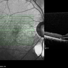
 APMPPE With Serous Macular Detachment SD-OCT
Jun 2 2014 by Rameez N Hussain, MD
SD OCT image of acute posterior multifocal placoid pigment epitheliopathy (APMPPE) with serous macular detachment.
Photographer: Rameez N Hussain MD, Vitreo Retinal Services, Giridhar Eye Institute, Cochin, India
Imaging device: Heidelberg Spectralis
Condition/keywords: acute posterior multifocal placoid pigment epitheliopathy (APMPPE), serous retinal detachment
-
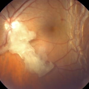 Retained Lens Fragment
Mar 2 2014 by Homayoun Tabandeh, MD, FASRS
Retained lens fragment, choroidal detachment, and serous retinal detachment post cataract surgery
Condition/keywords: retained lens fragments
-
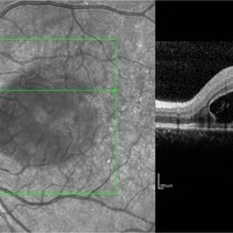
 APMPPE With Serous Macular Detachment 3D SD-OCT
Jun 2 2014 by Rameez N Hussain, MD
3D SD-OCT of acute posterior multifocal placoid pigment epitheliopathy (APMPPE) with serous macular detachment.
Photographer: Rameez N Hussain MD, Vitreo Retinal Services, Giridhar Eye Institute, Cochin, India
Imaging device: Heidelberg Spectralis
Condition/keywords: acute posterior multifocal placoid pigment epitheliopathy (APMPPE), serous retinal detachment
-
---thumb.jpg/image-square;max$300,300.ImageHandler) Choroidal Tumor, Breast Cancer
Choroidal Tumor, Breast Cancer
Feb 13 2013 by From the Collections of Thomas M. Aaberg, MD and Thomas M. Aaberg Jr., MD
Optic nerve, pt. LB, breast cancer, serous retinal detachment.
Condition/keywords: choroidal tumor, serous retinal detachment
-
 chronic central serous chorioretinopathy
Oct 31 2012 by Mallika Goyal, MD
Fluorescein angiogram of inferior retina of right eye with chronic CSCR shows dilation of and mild leak from retinal vessels over the inferior serous retinal detachment.
Condition/keywords: central serous chorioretinopathy (CSCR), chronic central serous chorioretinopathy (CSCR), serous retinal detachment
-
 Coats Disease
Sep 13 2013 by Maria Ana Martinez-Castellanos, MD
Peripheral fundus angiogram in a 2-years-old boy with Coat's disease.
Photographer: Maria A. Martinez-Castellanos. Asociacion para Evitar la Ceguera en Mexico
Imaging device: RetCAm II
Condition/keywords: pediatic retina, serous retinal detachment, vascular anomaly, vascular occlusions
-
---thumb.jpg/image-square;max$300,300.ImageHandler) Choroidal Tumor, Breast Cancer
Choroidal Tumor, Breast Cancer
Feb 13 2013 by From the Collections of Thomas M. Aaberg, MD and Thomas M. Aaberg Jr., MD
Optic nerve, pt. LB, breast cancer, serous retinal detachment.
Condition/keywords: choroidal tumor, serous retinal detachment
-
 Serous Retinal Detachment in Coats Disease
Mar 31 2014 by Maria Ana Martinez-Castellanos, MD
Fundus photograph of a 3-year-old boy with low vision, esotropia and leukocoria.
Photographer: Maria A. Martinez-Castellanos. Asociacion para Evitar la Ceguera en Mexico
Imaging device: RetCam II
Condition/keywords: pediatic retina, vascular anomaly
-
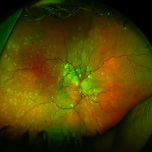 Hypertensive Retinopathy, Right
Feb 23 2017 by Alla Goldberg, MD
Fundus photograph of 35-year-old man with severe hypertension (182/128).
Photographer: Sofia Rutiaga, UT Health McGovern Medical School, Cizik Eye Clinic
Condition/keywords: cotton wool spots, Elschnig's spots, hypertensive choroidopathy, hypertensive retinopathy, serous retinal detachment
-
 Choroidal Tumor, Breast Cancer
Feb 13 2013 by From the Collections of Thomas M. Aaberg, MD and Thomas M. Aaberg Jr., MD
Optic nerve, pt. LB, breast cancer, serous retinal detachment.
Condition/keywords: serous retinal detachment
-
 Coloboma
Sep 7 2018 by John S. King, MD
11-year-old white female with bilateral optic nerve and retinochoroidal colobomas and an optic nerve pit in the right eye looking almost like pseudoduplication of the optic nerve. She is currently 20/30 OD and 20/20 OS. She has a history of laser by Dr. Zocchi about 10 years ago for a low lying, macula involving, serous retinal detachment, and has responded well.
Photographer: Stacey Coleman
Imaging device: Topcon
Condition/keywords: chorioretinal coloboma, inferior optic nerve coloboma, optic disc pit
-
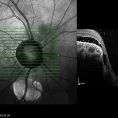
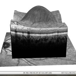
 Optic Pit
Jul 13 2016 by PAVEL FLORES-MORENO
OCT of a 56-year-old male with 7 days of low visual acuity.
Photographer: Flores-Moreno Pavel
Condition/keywords: optic pit, serous retinal detachment
-
---thumb.JPG/image-square;max$300,300.ImageHandler) chronic central serous chorioretinopathy
chronic central serous chorioretinopathy
Oct 31 2012 by Mallika Goyal, MD
Inferior serous retinal detachment in an eye with chronic CSCR.
Photographer: Mallika Goyal, MD
Condition/keywords: chronic central serous chorioretinopathy (CSCR)
 Loading…