Search results (73 results)
-
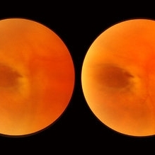
 Congenital Meridional
Nov 9 2012 by Norman Byer
This is the same case as seen in the previous photograph but showing an area just below the lower end of the dialysis. It shows a congenital meridional fold at the 2 o’clock meridian with a retinal break at the posterior end possibly caused by the direct injury described previously.
Condition/keywords: meridional fold, ora serrata, retinal break
-
 Enclosed Ora Bay On The Temporal Side
Nov 9 2012 by Norman Byer
This is a developmental abnormality in a 59-year-old man. It is an enclosed ora bay on the temporal side, an isolated island of normal pars plana epithelium. It is important not to confuse this entity with a retinal break. It has smooth, sloping borders not a sharp, thin, visible retinal edge as a retinal break would have. The border looks exactly like that of the ora serrata, and the grayish pigmented base has the same appearance as the normal pars plana.
Condition/keywords: developmental abnormality, enclosed ora bay, grayish pigmented base, horizontal nasal meridian, pars plana epithelium, smooth sloping borders, temporal retina
-
 Retinal Break at Site of Lattice Degeneration with Scleral Indentation
Nov 9 2012 by Norman Byer
This is the same case as the previous photograph. With scleral indentation slightly more posterior, the flap is seen to be associated with a large retinal tear. This is a tractional tear and it is possible that in this case the cryotherapy itself may have increased the vitreoretinal traction at this site and in this way led to this new tear. The age of the tear is unknown because it was asymptomatic, and even though the eye is aphakic the tear has not caused a clinical retinal detachment.
Condition/keywords: retinal flap, scleral indentation, tractional retinal tear, vitreoretinal traction
-
 Horseshoe Tear
Nov 9 2012 by Norman Byer
This horseshoe tear was the cause of the detachment in this 54-year-old man. The orange area on the right half of the slide represents the area of scleral indentation. Please note that most of the tear lies over the indented area and appears orange. However, the extreme left side of the tear is brownish black in color because it is exactly superimposed over the dark shadow that always lies just beyond the indented area. The ability of scleral indentation to produce this color change combined with a sharp demarcation between the blackish area and the yellowish edge of intact retina is a pathognomonic sign of a full thickness retinal break.
Condition/keywords: scleral indentation
-
 Pseudo Retinal Break
Nov 9 2012 by Norman Byer
This 23-year-old man presented with a fresh retinal detachment in a highly myopic eye and this very unusual retinal appearance. You can see two reddish areas with fairly distinct borders which at first make us think of retinal breaks. However, the left area has two tiny vessels visible in it, and the right area shows visible translucent retinal tissue extending across it. This patient has extensive areas of paving stone degeneration. Usually, such lesions present a barrier to a detaching retina and areas of paving stone usually remain attached. However, in this photograph we can see two paving stone lesions, and the detachment has extended right through them peeling them off from the underlying pigment epithelium. The two reddish areas, therefore, represent the very thin retina which previously constituted part of two paving stone lesions. The yellow atrophic areas which are visible deep to the detached retina represent the deeper parts of the same two original paving stone lesions.
Condition/keywords: lesion, myopic eye, pigment epithelium, reddish lesion, yellow atrophic area
-
 Retinal Schisis Detachment
Nov 9 2012 by Norman Byer
This 57-year-old man has a combined retinal schisis detachment caused by an outer layer hole in the upper right. On the right half of this photograph, the outer layer is detached and represented by the prominent yellow line which is lying against the inner layer. On the left half the inner layer appears very thin and the schisis cavity lies just behind it as it was originally. This, therefore, represents a localized detachment of the outer layer and thus a true secondary retinal detachment. The reason these cases remain localized and nonprogressive is that the only fluid available to the subretinal space is that which is contained within the schisis cavity. Furthermore, this fluid tends to be quite viscous and is not readily passed through the retinal breaks. A clinical symptomatic progressive retinal detachment cannot occur unless the retinal schisis cavity is very large or a break occurs in the inner layer also.
Condition/keywords: intact inner layer, localized detachment of outer layer, outer layer hole, retinal schisis detachment, retinoschisis, secondary retinal detachment
-
 Scleral Indentation
Nov 9 2012 by Norman Byer
This is the same lesion with scleral indentation. You can see the small discrete preretinal hemorrhage and the sharply circumscribed area of elevated retina with subretinal fluid beneath it. No retinal break is visible, but the posterior vitreous is detached and exerting traction at this site. The area was surrounded with argon laser treatment the same day as the initial examination.
Condition/keywords: posterior vitreous detachment, preretinal hemorrhage, scleral indentation, subretinal fluid, vitreous traction
-
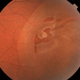
 Elevated Lesion
Nov 9 2012 by Norman Byer
This photograph and the next are two views of a very interesting elevated lesion in a 45-year-old man. This photograph shows the immense value of closely scrutinizing the profile of the indented area. Note that in the middle of the slide there is a sudden break in the continuity of the dark convex shadow that lies just behind the crest of the scleral indentation. If the elevated tissue is "filmy" or "wispy" or filamentous as in this case, it raises a strong suspicion that a retinal break is present just behind it.
Condition/keywords: elevated retinal lesion, elevated tissue, retinal break, scleral indentation
-
 Retinal Break
Nov 9 2012 by Norman Byer
This is the right eye of a 49-year-old woman showing a tiny retinal break adjacent to the temporal ora serrata. It has remained exactly the same without treatment for nine years.
Condition/keywords: ora serrata, retinal break
-
 Full-thickness Macular Hole
Aug 28 2012 by Sharon Fekrat, MD FACS FASRS
65 year old woman with a recurrent full thickness macular hole following previous 20 g pars plana vitrectomy in the right eye as well as an iatrogenic retinal hole in the papillomacular bundle. Both retinal defects are captured here in this Zeiss Stratus OCT image.
Photographer: Michael P. Kelly, FOPS Director, Duke Eye Labs, Duke University Eye Center, Durham, NC
Imaging device: Zeiss Cirrus
Condition/keywords: retinal break
-
 Pseudo Retinal Break
Nov 9 2012 by Norman Byer
The next three photographs will illustrate retinal conditions that can easily be mistaken for retinal breaks. For a fourth example of a pseudo retinal break, see slide pair 35. It is important to distinguish these conditions from true retinal breaks. This 49-year-old man was found to have this crescent shaped reddish lesion with a sharp yellow posterior border but without any visible elevated retinal flap. The two blood vessels which traversed this lesion in the presence of a flat retina proved that the retina is intact. This confusing appearance is caused by the presence of white with pressure both behind and in front of the reddish area causing it to resemble a retinal break.
Condition/keywords: pseudo retinal break, reddish lesion, retinal flap, traversing retinal vessels, white with pressure
-
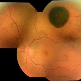
---thumb.JPG/image-square;max$300,300.ImageHandler) Retinoschisis
Retinoschisis
Oct 26 2012 by Mallika Goyal, MD
Fundus photograph of left eye of a 9-year-old boy with juvenile retinoschisis with large inner retinal break .
Condition/keywords: juvenile retinoschisis, retinal break
-
 Pseudo Retinal Break
Nov 9 2012 by Norman Byer
This large reddish area in the nasal periphery of this left eye is actually an enclosed ora bay. For other examples of this, see slide pair 5 and 6. This developmental abnormality could easily be confused with a retinal break. A very unusual feature of this photograph is the presence of a tiny true retinal break at the far right end of the enclosed ora bay and lying just to the left of the yellow zone in the middle of the photograph
Condition/keywords: enclosed ora bay, pseudo retinal break, reddish areas
-
---thumb.JPG/image-square;max$300,300.ImageHandler) Retinoschisis
Retinoschisis
Oct 26 2012 by Mallika Goyal, MD
Fundus photograph of left eye of a 9-year-old boy with juvenile retinoschisis with large inner retinal break.
Condition/keywords: juvenile retinoschisis, retinal break
-
 Retinal Detachment with Proliferative Vitreoretinopathy
Mar 20 2014 by Min Kim, MD, PhD, MBA, FASRS
Wide field fundus photograph of a 59-year-old male with chronic total RD and PVR, with multiple retinal breaks that developed a few months after LASIK surgery.
Photographer: Young Duk Bae, Yonsei University, Gangnam Severance Hospital
Imaging device: Wide field fundus photography, Optomap
Condition/keywords: proliferative vitreoretinopathy (PVR), retinal detachment with retinal defect
-
 Retinal Detachment Due to Traumatic Retinal Breaks
Mar 21 2013 by Yusuke Oshima, MD, PhD
Focal retinal detachment secondary to traumatic retinal breaks.
Photographer: Yusuke Takada, Osaka University Graduate School of Medicine
-
 CHRPE
CHRPE
Oct 8 2019 by DIEGO TOLENTINO
CHRPE plus laser barricade around retinal break
Photographer: Diego Tolentino
Condition/keywords: congenital hypertrophy of the retinal pigment epithelium (CHRPE)
-
 Retinal Fold Near Retinal Break in Rhegmatogenous Retinal Detachment
Dec 15 2014 by Mallika Goyal, MD
Left fundus of a 32-year-old male shows a fixed retinal fold. This is adjacent to a large retinal break (not seen here) with rhegmatogenous retinal detachment.
Photographer: Mallika Goyal, MD, Apollo Health City, Jubilee Hills, Hyderabad-500033
Condition/keywords: retinal fold
-
 Bilateral Senile Retinoschisis
Apr 22 2016 by Mallika Goyal, MD
Left fundus of a 70-year-old male with bilateral asymptomatic bullous inferotemporal senile retinoschisis. There were no inner or outer layer retinal breaks either eye.
Photographer: Mallika Goyal, MD, Apollo Health City, Hyderabad, India
Condition/keywords: senile retinoschisis
-
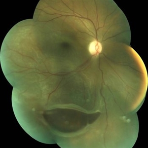
 Asymptomatic Superior Retinal Detachment
May 5 2016 by Steven J Ryder, MD
38-year-old African American female with moderate myopia (-4.50 Sph OU) and asymptomatic superior retinal detachment in the right eye. Montage fundus photography showing localized retinal detachment superiorly with single full-thickness retinal break at 12:00.
Photographer: Luis Bernhard, Miami VA Healthcare System
Imaging device: Topcon
Condition/keywords: asymptomatic, full thickness retinal hole, myopia, retinal break, retinal detachment with retinal defect
-
 Retinal Detachment
Apr 18 2014 by Neha Goel, MS DNB FRCS (Glasg)
Fundus photograph of a 35-year-old female with subtotal retinal detachment and a large inferior, posterior break.
Photographer: Kiran Sharma, Guru Nanak Eye Centre, Maulana Azad Medical College, New Delhi, India
Imaging device: Zeiss Visucam
Condition/keywords: posterior break, retinal break
-
 Surgical Displacement of Subfoveal Subretinal Hemorrhage Using rt-PA, Postop Day One
Oct 15 2012 by Sharon Fekrat, MD FACS FASRS
Fundus photograph of a 76 year old female with neovascular AMD who developed a subfoveal subretinal hemorrhage in the left eye. This photograph is one day postop after 23g vitrectomy, subretinal rt-PA 50ug/0.1cc and C3F8 gas. Note the subretinal hemorrhage and fluid displaced inferiorly. No open retinal break was present.
Photographer: Duke University Eye Center, Durham, NC
-
---thumb.jpg/image-square;max$300,300.ImageHandler) Rhegmatogeous Retinal Detachment
Rhegmatogeous Retinal Detachment
Mar 21 2013 by Yusuke Oshima, MD, PhD
Wide-field fundus photograph of a 58-year-old woman with a macula-involved bullous retinal detachment due to a superotemporal retinal break.
Photographer: Yusuke Takada, Osaka University Graduate School of Medicine
Imaging device: OPTOS 200Tx
Condition/keywords: bullous retinal detachment
-
 Bilateral Senile Retinoschisis
Apr 22 2016 by Mallika Goyal, MD
Left fundus of a 70-year-old male with bilateral asymptomatic bullous inferotemporal senile retinoschisis. There were no inner or outer layer retinal breaks either eye.
Photographer: Mallika Goyal, MD, Apollo Health City, Hyderabad, India
Condition/keywords: senile retinoschisis
-
 Retinal Fold Near Retinal Break in Rhegmatogenous Retinal Detachment
Dec 15 2014 by Mallika Goyal, MD
Left fundus of a 32-year-old male shows a fixed retinal fold near a large retinal break with rhegmatogenous retinal detachment.
Photographer: Mallika Goyal, MD, Apollo Health City, Jubilee Hills, Hyderabad-500033
Condition/keywords: retinal fold
 Loading…