 Initializing download.
-
 By Norman Byer
From Dr. Norman E. Byer’s “The Peripheral Retina in Profile” - Uploaded on Nov 9, 2012.
- Last modified by Suber S. Huang, MD, MBA, FASRS on Feb 11, 2013.
- Reviewed by Chayal Patel
- Rating
- Appears in
- Miscellaneous
- Condition/keywords
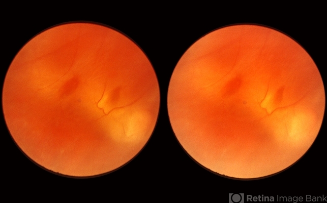
- myopic eye, reddish lesion, yellow atrophic area, pigment epithelium
- Description
- This 23-year-old man presented with a fresh retinal detachment in a highly myopic eye and this very unusual retinal appearance. You can see two reddish areas with fairly distinct borders which at first make us think of retinal breaks. However, the left area has two tiny vessels visible in it, and the right area shows visible translucent retinal tissue extending across it. This patient has extensive areas of paving stone degeneration. Usually, such lesions present a barrier to a detaching retina and areas of paving stone usually remain attached. However, in this photograph we can see two paving stone lesions, and the detachment has extended right through them peeling them off from the underlying pigment epithelium. The two reddish areas, therefore, represent the very thin retina which previously constituted part of two paving stone lesions. The yellow atrophic areas which are visible deep to the detached retina represent the deeper parts of the same two original paving stone lesions.


