Search results (65 results)
-
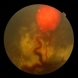
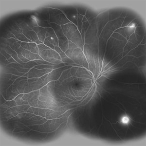
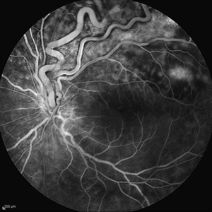
 Retinal Angiomatous Proliferation in Age-Related Macular Degeneration with Subretinal Neovascularization
Sep 24 2012 by James B. Soque, CRA, OCT-C, COA, FOPS
75-year-old white male with classic SRN with RAP. Lesion OD is active, and patient is receiving anti-VEGF treatment. Mid phase FA, 50 Deg, Mag 2x.
Photographer: James Soque, CRA, COA, Island Retina, Shirley, NY, USA
Imaging device: Topcon TRC 50 DX, OIS 5.0 MP Color, FA Camera, OIS Software
Condition/keywords: age-related macular degeneration (AMD), fundus autofluorescence (FAF), leakage, retinal angiomatous proliferation (RAP), subretinal neovascularization (SRNV)
-
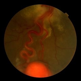
 Von Hippel-Lindau
Sep 3 2012 by Hamid Ahmadieh, MD
Color fundus photograph of a 35-year-old woman with retinal angiomatosis.
Photographer: Hamid Ahmadieh, MD, Ophthalmic Research Center, Labbafinejad Medical Center, Shahid Beheshti University of Medical Sciences , Tehran
Imaging device: Topcon Fundus Camera
Condition/keywords: retinal angiomatous proliferation (RAP), Von Hippel-Lindau
-
 Von Hippel-Lindau
Sep 3 2012 by Hamid Ahmadieh, MD
Color fundus photograph of a 35-year-old woman with retinal angiomatosis.
Photographer: Hamid Ahmadieh, MD, Ophthalmic Research Center, Labbafinejad Medical Center, Shahid Beheshti University of Medical Sciences
Imaging device: Topcon Fundus Camera
Condition/keywords: retinal angiomatous proliferation (RAP), Von Hippel-Lindau
-
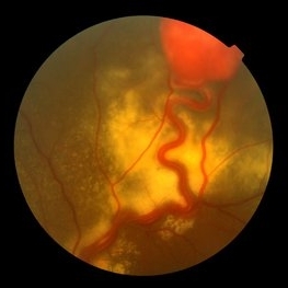
 Retinal Angiomas In VHL
Dec 24 2012 by Roy D. Brod, MD
Fundus photograph of 16 year old male with recent diagnosis of Von Hippel-Lindau disease showing typical appearance of a retinal angioma in superior mid periphery OD. Note unrelated choroidal nevus above superior arcade.
Photographer: Julia Walker
Condition/keywords: hemangioma, Von Hippel-Lindau
-
 Von Hippel-Lindau
Sep 5 2012 by Hamid Ahmadieh, MD
Color fundus photograph of a 32-year-old man with retinal angiomatosis.
Photographer: Hamid Ahmadieh, MD, Ophthalmic Research Center, Labbafinejad Medical Center, Shahid Beheshti University of Medical Sciences
Imaging device: Topcon Fundus Camera
Condition/keywords: retinal angiomatous proliferation (RAP), Von Hippel-Lindau
-
 Von Hippel-Lindau
Sep 8 2012 by Hamid Ahmadieh, MD
Color fundus photograph of a 32-year-old man with retinal angiomatosis.
Photographer: Hamid Ahmadieh, MD, Ophthalmic Research Center, Labbafinejad Medical Center, Shahid Beheshti University of Medical Sciences
Imaging device: Topcon Fundus Camera
Condition/keywords: retinal angiomatous proliferation (RAP), Von Hippel-Lindau
-
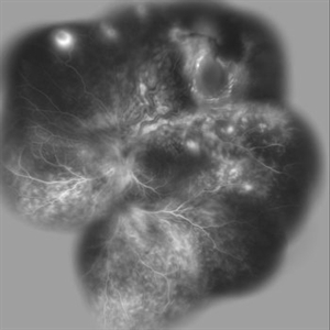
 Von Hippel-Lindau
Oct 13 2012 by Hamid Ahmadieh, MD
Wide field FA image of the right eye of a 25-year-old woman with retinal angiomatosis (Von Hippel-Lindau). Fundus of the right eye seemed to be normal in ophthalmoscopy.
Photographer: Soodabeh Fooladin, Negah Eye Center, Tehran
Imaging device: Heidelberg Spectralis
Condition/keywords: exudative retinal detachment, retinal angiomatous proliferation (RAP), Von Hippel-Lindau
-
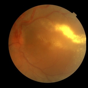 Von Hippel-Lindau 1
Oct 13 2012 by Hamid Ahmadieh, MD
Color fundus photograph of the left eye of a 25-year-old woman with exudative retinal detachment secondary to retinal angiomatosis (Von Hippel-Lindau).
Photographer: Hamid Ahmadieh, MD, Ophthalmic Research Center, Labbafinejad Medical Center, Shahid Beheshti University of Medical Sciences
Imaging device: Topcon Fundus Camera
Condition/keywords: exudative retinal detachment, retinal angiomatous proliferation (RAP), Von Hippel-Lindau
-
 Von Hippel-Lindau
Sep 3 2012 by Hamid Ahmadieh, MD
Color fundus photograph of a 35-year-old woman with retinal angiomatosis.
Photographer: Hamid Ahmadieh, MD, Ophthalmic Research Center, Labbafinejad Medical Center, Shahid Beheshti University of Medical Sciences
Imaging device: Topcon Fundus Camera
Condition/keywords: retinal angiomatous proliferation (RAP)
-
 Retinal Angiomas In VHL
Dec 24 2012 by Roy D. Brod, MD
Fundus photograph of 16 year old male with recent diagnosis of Von Hippel-Lindau disease showing 2 retinal angiomas in inferior mid periphery OD.
Photographer: Julia Walker
Condition/keywords: hemangioma, Von Hippel-Lindau
-
 Von Hippel-Lindau
Oct 13 2012 by Hamid Ahmadieh, MD
Wide field FA image of the left eye of a 25-year-old woman with exudative retinal detachment secondary to retinal angiomatosis (Von Hippel-Lindau).
Photographer: Soodabeh Fooladin, Negah Eye Center, Tehran
Imaging device: Heidelberg Spectralis
Condition/keywords: exudative retinal detachment, retinal angiomatous proliferation (RAP), Von Hippel-Lindau
-
 Von Hippel-Lindau
Oct 13 2012 by Hamid Ahmadieh, MD
Late FA image of the left eye of a 25-year-old woman with exudative retinal detachment secondary to retinal angiomatosis (Von Hippel-Lindau).
Photographer: Soodabeh Fooladin, Negah Eye Center, Tehran
Imaging device: Heidelberg Spectralis
Condition/keywords: exudative retinal detachment, retinal angiomatous proliferation (RAP)
-
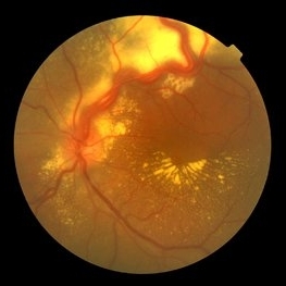
 Retinal Angiomatous Proliferation
Oct 11 2012 by Gabriela Lopezcarasa Hernandez, MD
70-year-old male with diagnostic of RAP by OCT and ICG fluorescein angiography
Photographer: Azucena Rios, Macula Retina Consultores Mexico
Imaging device: Heidelberg Spectralis
Condition/keywords: retinal angiomatous proliferation (RAP)
-
 Choroidal Neovascularization with Retinal Angiomatous Proliferation
Aug 24 2012 by John S. King, MD
3.35 min
Photographer: Kristin Konecki, OcuSight Eye Care Center, Rochester, NY
Condition/keywords: retinal angiomatous proliferation (RAP)
-
 Choroidal Neovascularization with Retinal Angiomatous Proliferation
Aug 24 2012 by John S. King, MD
IRH in area of drusen both juxtafoveal and extrafoveal. Photo and FAs are initial presentation
Photographer: Kristin Konecki, OcuSight Eye Care Center, Rochester, NY
Condition/keywords: retinal angiomatous proliferation (RAP)
-
 Choroidal Neovascularization with Retinal Angiomatous Proliferation
Aug 24 2012 by John S. King, MD
Before and 1 week post Avastin; PED, SRF, ME.
Photographer: Kristin Konecki, OcuSight Eye Care Center, Rochester, NY
Condition/keywords: Avastin, retinal angiomatous proliferation (RAP)
-
 Retinal Angiomatous Proliferation
Jan 29 2017 by ADRIANO FERREIRA
Fundus photograph of an 56-year-old man with a retinal angiomatous proliferation (RAP). RAP has been known as a variant of exudative age-related macular degeneration (AMD) with a unfavorable prognosis.
Photographer: Laercio
Condition/keywords: vascular anomaly
-
 Choroidal Neovascularization with Retinal Angiomatous Proliferation
Aug 24 2012 by John S. King, MD
16 sec
Photographer: Kristin Konecki, OcuSight Eye Care Center, Rochester, NY
Condition/keywords: retinal angiomatous proliferation (RAP)
-
 Choroidal Neovascularization with Retinal Angiomatous Proliferation
Aug 24 2012 by John S. King, MD
1.02 min
Photographer: Kristin Konecki, OcuSight Eye Care Center, Rochester, NY
Condition/keywords: retinal angiomatous proliferation (RAP)
-
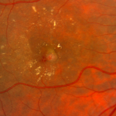
 Retinal Angioma With Lipid In The Fovea
Jul 11 2013 by Jerald A. Bovino, MD
Probably is post-rx.
Condition/keywords: fovea, lipid, retinal angioma
-
 Retinal Angiomatosis in a 21-Year-Old Male - 2
Aug 11 2015 by Roy Schwartz, MD
Fundus photograph of a 21-year-old man, who on routine examination was found to have two capillary hemangioblastomas in his left eye. He was diagnosed with retinal angiomatosis.
Photographer: Galit Yair Pur
Condition/keywords: retinal angioma
-
 Retinal Angioma With Lipid In The Fovea
Jul 11 2013 by Jerald A. Bovino, MD
Probably is pre-rx.
Condition/keywords: fovea, lipid, retinal angioma
-
 Retinal Angiomatosis in a 21-Year-Old Male - 1
Aug 11 2015 by Roy Schwartz, MD
Fundus photograph of a 21-year-old man, who on routine examination was found to have two capillary hemangioblastomas in his left eye. He was diagnosed with retinal angiomatosis.
Photographer: Galit Yair Pur
Condition/keywords: retinal angioma
-
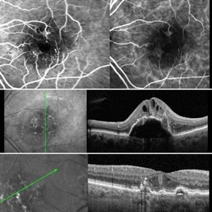 Retinal Angiomatous Proliferation RAP
Mar 11 2020 by RAFAEL REIS PEREIRA, MD
Retinal angiomatous proliferation (RAP) is a unique variant of neovascular age-related macular degeneration. Published studies have estimated that up to 15% of patients with neovascular age-related macular degeneration have RAP. Clinical features frequently associated with RAP include bilateral disease, presence of pigment epithelial detachments, and reticular pseudodrusen. RAP is more frequently associated with the development of retinal pigment epithelial tears and geographic atrophy that can lead to severe vision loss. We present a stereo fluorescein angiography and ICG (upper right and left image respectively) and OCT of left and right eye (middle and inferior image) of a RAP choroidal neovascularization in an 89-year-old patient.
Photographer: Rafael Reis Pereira
Imaging device: HRA Heildelberg Spectralis
Condition/keywords: retinal angiomatous proliferation (RAP)
-
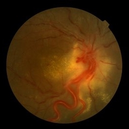
 Stereo View of Retinal Angiomatous Proliferation in Age-Related Macular Degeneration
Jan 21 2016 by James B. Soque, CRA, OCT-C, COA, FOPS
Stereo pair of 75-year-old white male with classic SRN with RAP lesion of right eye, actively receiving anti-VEGF treatment. 50 Degrees, no mag, L and R stereo pair. Single View of OD also visible in this case.
Condition/keywords: age-related macular degeneration (AMD), anti-VEGF, retinal angiomatous proliferation (RAP), stereo pair, subretinal neovascularization (SRNV)
 Loading…