Search results (66 results)
-
 Uveitis Posterior
Jul 19 2019 by JEFFERSON R SOUSA, Tecg.º (Biomedical Systems Technology)
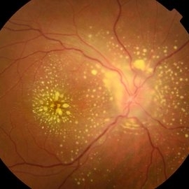
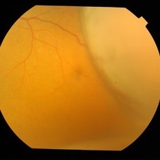
A 23-year-old male patient attended the clinic with low vision of the right eye. In the evaluation it presented important fundoscopical alterations like retinal exudations in the posterior pole and nasal retina, aspects of macular star. It was proven that it was a posterior uveitis.
Photographer: JEFFERSON R SOUSA - Study Center and Ophthalmological Research Dr. Andre M V Gomes, Institute Dr. Suel Abujamra São Paulo-Brazil
Imaging device: Topcon TRC-50 DX, Imaginet 4.0, angle de 50 graus. Flash 50w-s
Condition/keywords: uveitis
-
 BSC CME OS
Nov 10 2012 by Pauline T Merrill, MD, FASRS
Fundus photograph left eye of a 42-year-old Caucasian male with birdshot retinochoroidopathy (HLA-A29+) and cystoid macular edema (CME)
Condition/keywords: birdshot retinochoroidopathy, cystoid macular edema (CME), posterior uveitis, uveitis
-
 Toxoplasmosis Slide 1
Oct 22 2012 by Ronald C. Gentile, MD
Focal, white area of chorioretinitis with overlying vitreous inflammation adjacent to an old chorioretinal scar in a patient complaining of photophobia, floaters and a decrease in vision of the right eye. The focal area of chorioretinitis is involving the inferior nasal macula and adjacent optic nerve with surrounding retinal and peri-papillary edema.
Photographer: The New York Eye & Ear Infirmary Department of Medical Imaging
Condition/keywords: posterior uveitis, toxoplasmosis
-
 Toxoplasmosis Slide 2
Oct 22 2012 by Ronald C. Gentile, MD
One month following treatment with Bactrim, Clindamycin, and oral prednisone the focal area chorioretinitis has coalesced with a decrease in overlying vitreous inflammation. Kyrieleis plaques can be seen along the inferior retinal arteriole.
Photographer: The New York Eye & Ear Infirmary Department of Medical Imaging
Condition/keywords: posterior uveitis, toxoplasmosis
-
---thumb.jpg/image-square;max$300,300.ImageHandler) Posterior Uveitis
Posterior Uveitis
Feb 15 2013 by From the Collections of Thomas M. Aaberg, MD and Thomas M. Aaberg Jr., MD
Color photograph of the mid-peripheral retina showing scattered intraretinal hemorrhage and foci of retinal whitening consistent with posterior uveitis, such as Behcet disease.
Condition/keywords: posterior uveitis, retinitis
-
 Uveitis With Exudative Retinal Detachment
May 3 2014 by Mallika Goyal, MD
Fluorescein angiogram of an elderly patient with bilateral posterior uveitis shows punctate hyperfluorescence and inferior RD. He responded well to oral steroids with complete resolution of the uveitis and RD.
Photographer: Mallika Goyal, MD, Apollo Health City, Jubilee Hills, Hyderabad, India
Condition/keywords: exudative retinal detachment, uveitis
-
---thumb.jpg/image-square;max$300,300.ImageHandler) Behcet Uveitis
Behcet Uveitis
Feb 15 2013 by From the Collections of Thomas M. Aaberg, MD and Thomas M. Aaberg Jr., MD
Color fundus photographs of the right eye of a patient suspected to have Behcet Uveitis. Over the course of 11 days, there is progressive optic disc edema, intraretinal whitening, hemorrhage and vessel occlusion.
Condition/keywords: Behcet's uveitis, posterior uveitis, retinitis
-
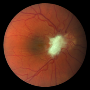 Pre-Retinal Fibrosis Following Endogenous Fungal Endophthalmitis
Aug 23 2018 by Matthew Dombrow, MD
20-year-old male presents with posterior uveitis with a chain of white infiltrates stemming from the optic nerve. While treated, his infiltrates retracted and hemorrhage occured. Neovascularization of the disc developed and underwent Avastin treatment in addition to his oral anti-fungals and intravitreal anti-fungals. Patient was lost to follow up and presented with severe pre-retinal fibrosis 6 years later. Acuity is 20/40-1 with significant metamorphopsia.
Photographer: Patricia Candrea, COA, Connecticut Retina Consultants, LLC
Imaging device: Canon
Condition/keywords: pre-retinal membrane
-
 Uveitis With Exudative Retinal Detachment
May 3 2014 by Mallika Goyal, MD
Fluorescein angiogram of an elderly patient with bilateral posterior uveitis shows punctate hyperfluorescence and inferior RD. He responded well to oral steroids with complete resolution of the uveitis and RD.
Photographer: Mallika Goyal, MD, Apollo Health City, Jubilee Hills, Hyderabad, India
Condition/keywords: exudative retinal detachment, uveitis
-
 Retinal Dialysis (Superior) and Macula Off Detachment
Jan 23 2017 by Nelson Chamma Capelanes, MD
Retinal dialysis (superior) and macula off detachment after toxoplasmosis posterior uveitis.
Photographer: Nelson Chamma Capelanes, Promedica Indaiatuba
Condition/keywords: retinal dialysis
-
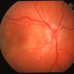 Posterior Uveitis
Apr 8 2019 by Gary R. Cook, MD, FACS
37-year-old white male with mild vitritis, optic disc hyperemia and edema, peripapillary hemorrhages and yellow-white spots in temporal macula OD; V.A. = 20/30.
Imaging device: Topcon VT-50
Condition/keywords: posterior uveitis
-
---thumb.jpg/image-square;max$300,300.ImageHandler) Posterior Uveitis
Posterior Uveitis
Feb 15 2013 by From the Collections of Thomas M. Aaberg, MD and Thomas M. Aaberg Jr., MD
Schematic diagram depicting Posterior Uveitis characterized by diffuse retinal whitening and exudative retinal detachment.
Condition/keywords: posterior uveitis, retinitis
-
---thumb.jpg/image-square;max$300,300.ImageHandler) Posterior Uveitis
Posterior Uveitis
Feb 15 2013 by From the Collections of Thomas M. Aaberg, MD and Thomas M. Aaberg Jr., MD
Reprint of schematic drawings depicting Posterior Uveitis showing acute retinal lesions and vitreous debris (left panel) that are eventually replaced by areas of well-defined retinal pigment epithelial atrophy (right panel).
Condition/keywords: posterior uveitis, retinitis
-
---thumb.jpg/image-square;max$300,300.ImageHandler) Behcet Uveitis
Behcet Uveitis
Feb 15 2013 by From the Collections of Thomas M. Aaberg, MD and Thomas M. Aaberg Jr., MD
Color fundus photographs of the right eye of a patient suspected to have Behcet Uveitis. Over the course of 11 days, there is progressive optic disc edema, intraretinal whitening, hemorrhage and vessel occlusion. Fluorescein angiography confirms impaired retinal perfusion secondary to vessel occlusion.
Condition/keywords: posterior uveitis, retinitis
-
 Retinal Dialysis (Superior) and Macula Off Detachment
Jan 23 2017 by Nelson Chamma Capelanes, MD
Retinal dialysis (superior) and macula off detachment after toxoplasmosis posterior uveitis.
Photographer: Nelson Chamma Capelanes, Promedica Indaiatuba
Condition/keywords: retinal dialysis
-
---thumb.jpg/image-square;max$300,300.ImageHandler) Posterior Uveitis
Posterior Uveitis
Feb 15 2013 by From the Collections of Thomas M. Aaberg, MD and Thomas M. Aaberg Jr., MD
Schematic diagram depicting Posterior Uveitis characterized by diffuse retinal whitening and exudative retinal detachment.
Condition/keywords: posterior uveitis, retinitis
-
---thumb.jpg/image-square;max$300,300.ImageHandler) Behcet Disease
Behcet Disease
Feb 15 2013 by From the Collections of Thomas M. Aaberg, MD and Thomas M. Aaberg Jr., MD
Reprint of a fundus photograph from a patient with Behcet disease showing optic atrophy, vessel narrowing, and pigmentary changes (from Colvard et al, Arch Ophthalmol 1977;95(10):1813-7).
Condition/keywords: posterior uveitis, retinitis
-
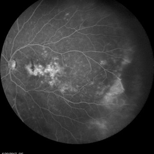 Posterior Uveitis with Cystoid Macular Edema
Jan 18 2018 by Olivia Rainey
Ultra-wide field fluorescein angiogram of a 59-year-old female with posterior uveitis and chronic cystoid macular edema affecting her left eye. Interestingly, she has peripheral capillary nonperfusion inferotemporal, which could be driving CME.
Photographer: Olivia Rainey
Imaging device: Heidelberg Spectralis
Condition/keywords: 102 degrees, cystoid macular edema (CME), fluorescein leakage, Heidelburg Spectralis, left eye, peripheral retinal nonperfusion, posterior uveitis, ultra-wide field imaging
-
 Retinal Dialysis (Superior) and Macula Off Detachment
Jan 23 2017 by Nelson Chamma Capelanes, MD
Retinal dialysis (superior) and macula off detachment after toxoplasmosis posterior uveitis.
Photographer: Nelson Chamma Capelanes, Promedica Indaiatuba
Condition/keywords: retinal dialysis
-
 Uveitis With Exudative Retinal Detachment
May 3 2014 by Mallika Goyal, MD
Early phase luorescein angiogram of an elderly patient with bilateral posterior uveitis shows punctate hyperfluorescence. He responded well to oral steroids with complete resolution of the uveitis.
Photographer: Mallika Goyal, MD, Apollo Health City, Jubilee Hills, Hyderabad, India
Condition/keywords: exudative retinal detachment, uveitis
-
---thumb.jpg/image-square;max$300,300.ImageHandler) Posterior Uveitis
Posterior Uveitis
Feb 15 2013 by From the Collections of Thomas M. Aaberg, MD and Thomas M. Aaberg Jr., MD
Progression of disease showing expansion and confluence of areas of retinal whitening over the course of 7 days.
Condition/keywords: posterior uveitis, retinitis
-
 Serpiginous Choroiditis
Sep 22 2019 by Haider Ali
35-year-old female presented with decrease in vision in her left eye for last 4 days and in right eye for last 8 days. Her right eye was previously involved in a similar episode about 5-6 months ago for which she was treated with oral steroids.
Photographer: Dr Haider Ali Chaudhry, Madinah Teaching Hospital, Faisalabad
Condition/keywords: acute posterior multifocal placoid pigment epitheliopathy (APMPPE), macula serpiginous choroidopathy, posterior uveitis, serpiginous choroiditis, uveitis, white dot lesions, white dot syndrome
-
 Uveitis With Exudative Retinal Detachment
May 3 2014 by Mallika Goyal, MD
Fundus photograph of an elderly patient with bilateral posterior uveitis and subnormal vision. Posterior pole looks relatively unremarkable though periphery reveal s choroidal detachments and exudative retinal detachment. Fluorescein angiogram reveals punctate hyperfluorecence through the entire fundus and disc hyperfluorescence.
Photographer: Mallika Goyal, MD, Apollo Health City, Jubilee Hills, Hyderabad, India
Condition/keywords: exudative retinal detachment, uveitis
-
 Serpiginous Choroiditis
Sep 22 2019 by Haider Ali
35-year-old female presented with decrease in vision in her left eye for last 4 days and in right eye for last 8 days. Her right eye was previously involved in a similar episode about 5-6 months ago for which she was treated with oral steroids.
Photographer: Dr Haider Ali Chaudhry, Madinah Teaching Hospital, Faisalabad
Condition/keywords: acute posterior multifocal placoid pigment epitheliopathy (APMPPE), macula serpiginous choroidopathy, posterior uveitis, serpiginous choroiditis, uveitis, white dot lesions, white dot syndrome
-
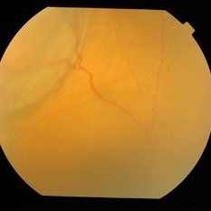
---thumb.JPG/image-square;max$300,300.ImageHandler) Birdshot Chorioretinopathy
Birdshot Chorioretinopathy
Jul 14 2013 by Jason S. Calhoun
Follow up on patient with birdshot chorioretinopathy in both eyes. Posterior uveitis in both eyes no changes in inflammation.
Photographer: Jason S. Calhoun, Department of Ophthalmology, Mayo Clinic Jacksonville, Florida
Imaging device: TOPCON TRC 50-EX
Condition/keywords: birdshot chorioretinopathy
 Loading…