Search results (63 results)
-
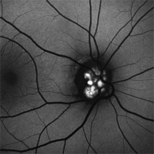
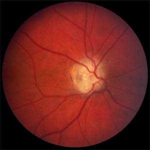
 Optic Disc Drusen
Jul 31 2016 by Mitzy E Torres Soriano, MD
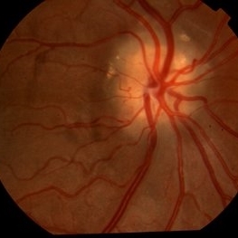
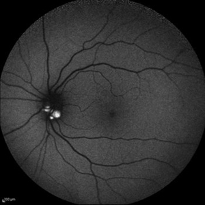
Optic Disc Drusen (Right eye)
Photographer: Mitzy E. Torres Soriano. Retina Department. Hospital Provincial del Centenario. Rosario, Argentina
Imaging device: TOPCON
Condition/keywords: optic disc drusen, optic nerve drusen
-
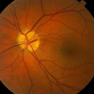
 Optic Nerve Head Drusen With Idiopathic CNV
Feb 17 2017 by Kristen Wagner
22-year-old female fundus photograph of a right eye with Optic Nerve Drusen with Idiopathic CNV.
Photographer: Kristen Wagner, COT, OSC Ophthalmic Photographer, Tennessee Retina, Nashville TN
Condition/keywords: choroidal neovascularization (CNV), drusen of optic disc, optic disc drusen
-
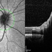
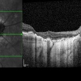
---thumb.jpg/image-square;max$300,300.ImageHandler) Optic Disc Drusen
Optic Disc Drusen
Jul 10 2013 by Hamid Ahmadieh, MD
SD-OCT image of the left eye of a 24-year-old woman with optic disc drusen and VA 20/20.
Photographer: Solmaz Shahmohammadi, Negah Eye Center, Tehran
Imaging device: Heidelberg Spectralis
Condition/keywords: optic disc drusen, optical coherence tomography (OCT)
-
 Optic Disc Drusen
Jul 10 2013 by Hamid Ahmadieh, MD
Fundus autofluorescence image of the right eye of a 24-year-old woman with optic disc drusen and VA 20/20.
Photographer: Solmaz Shahmohammadi, Negah Eye Center, Tehran
Imaging device: Heidelberg Spectralis
Condition/keywords: fundus autofluorescence (FAF), optic disc drusen
-
 Optic Disc Drusen Autofluorescence
Apr 2 2016 by David Callanan, MD
30-year-old Caucasian male with visual field defect OD > OS.
Condition/keywords: optic disc drusen
-
 Optic Disc Drusen
Sep 21 2012 by Suber S. Huang, MD, MBA, FASRS
Fundus photograph of a 50-year-old woman with optic disc drusen complicated by anterior ischemic optic neuropathy
Condition/keywords: optic disc drusen
-
 Buried drusen with CNV
Dec 19 2012 by Eric A. Postel, MD
Buried optic disc drusen complicated by peripapillary subretinal neovascularisation
Condition/keywords: choroidal neovascularization (CNV), optic disc drusen
-
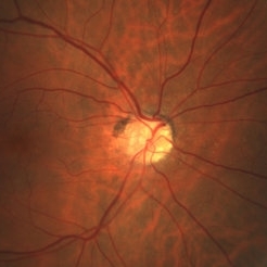
---thumb.jpg/image-square;max$300,300.ImageHandler) Optic Disc Drusen
Optic Disc Drusen
Mar 27 2013 by Henry J. Kaplan, MD
An 11-year-old boy presented with transient blurry vision, VA:20/20 bilaterally. He has pseudo optic disc swelling only in the right eye ; margin is blurred but the pattern of vessels are normal and there are some yellowish deposits on the superior of ON #1. AF, B-scan, CT scan, and VF are uploaded in the following slides.
Condition/keywords: drusen of optic disc, optic disc drusen, optic nerve drusen
-
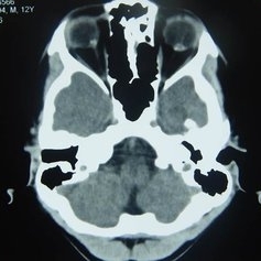 Orbital CT Scan in Optic Nerve Drusen
Mar 27 2013 by Henry J. Kaplan, MD
Axial CT scan of orbit demonstrates high density spot on optic nerve head on both sides #4.
Condition/keywords: drusen of optic disc, optic disc drusen, optic nerve drusen
-
 Optic Disc Drusen
Jul 10 2013 by Hamid Ahmadieh, MD
Fundus autofluorescence image of the left eye of a 24-year-old woman with optic disc drusen and VA 20/20.
Photographer: Solmaz Shahmohammad, Negah Eye Center, Tehran
Imaging device: Heidelberg Spectralis
Condition/keywords: fundus autofluorescence (FAF), optic disc drusen
-
 Optic disc drusen, left eye - L stereo
Jan 11 2013 by Alex P. Hunyor, MD
Optic disc drusen, left eye - L stereo.
Condition/keywords: drusen of optic disc, optic disc drusen
-
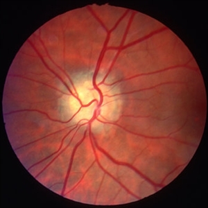 Optic disc drusen, left eye - R stereo
Jan 11 2013 by Alex P. Hunyor, MD
Optic disc drusen, left eye - R stereo.
Condition/keywords: drusen of optic disc, optic disc drusen
-
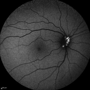
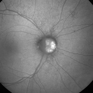 Optic disc drusen
Dec 25 2012 by Alex P. Hunyor, MD
Autofluorescent image of the right optic disc showing autofluorescence of optic disc drusen.
Condition/keywords: optic disc drusen
-
 Optic Disc Drusen
Jul 10 2013 by Hamid Ahmadieh, MD
SD-OCT image of the right eye of a 24-year-old woman with optic disc drusen and VA 20/20.
Photographer: Solmaz Shahmohammadi, Negah Eye Center, Tehran
Imaging device: Heidelberg Spectralis
Condition/keywords: optic disc drusen, optical coherence tomography (OCT)
-
 Optic Disc Drusen
Jul 10 2013 by Hamid Ahmadieh, MD
SD-OCT image of the right eye of a 24-year-old woman with optic disc drusen and VA 20/20.
Photographer: Solmaz Shahmohammadi, Negah Eye Center, Tehran
Imaging device: Heidelberg Spectralis
Condition/keywords: optic disc drusen, optical coherence tomography (OCT)
-
---thumb.jpg/image-square;max$300,300.ImageHandler) Optic Disc Drusen
Optic Disc Drusen
Mar 27 2013 by Henry J. Kaplan, MD
Autofluorescence imaging shows heper AF on the optic nerve head specially superiorly due to drusen in the same patient #2.
Imaging device: Heidelberg spectralis
Condition/keywords: drusen of optic disc, optic disc drusen, optic nerve drusen
-
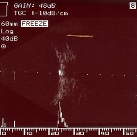 Optic Disc Drusen
Mar 27 2013 by Henry J. Kaplan, MD
B-scan of the same patient with low gain (48db), demonstrates high spike on the optic nerve head due to drusen #3.
Condition/keywords: drusen of optic disc, optic disc drusen, optic nerve drusen
-
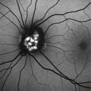
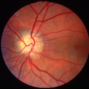
 Optic disc drusen
Dec 25 2012 by Alex P. Hunyor, MD
Colour image showing right optic disc drusen.
Condition/keywords: optic disc drusen
-
 Optic Disc Drusen Autofluorescence
Apr 2 2016 by David Callanan, MD
30-year-old Caucasian male with visual field defect OD > OS.
Condition/keywords: optic disc drusen
-
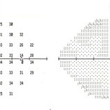 Optic Disc Drusen
Mar 27 2013 by Henry J. Kaplan, MD
Perimetry demonstrates slightly enlarged blind spot in the same patient #5.
Condition/keywords: drusen of optic disc, optic disc drusen, optic nerve drusen
-
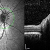
 Optic Nerve Head Drusen with OCT
Feb 2 2018 by Olivia Rainey
Optical coherence tomography with enhanced depth imaging of a 86-year-old male with optic nerve head drusen affecting his right eye. This patient has also been diagnosed with pseudoxanthoma elasticum and macular degeneration.
Photographer: Olivia Rainey
Imaging device: Heidelberg Spectralis
Condition/keywords: enhanced depth imaging, infrared image, macular degeneration, optic disc drusen, optic nerve, optical coherence tomography (OCT), pseudoxanthoma elasticum (PXE)
-
 Color Fundus Photographs of Optic Disc Drusen
Apr 26 2018 by Ahmad B. Tarabishy, MD
Fundus photographs and autofluorescence of a 75-year-old man with an epiretinal membrane in the left eye. Incidentally, he had a history of optic disc drusen, which show a striking hyperautofluorescence on FAF imaging.
Photographer: Michelle Howarth, Lakeland Eye Clinic
Imaging device: Zeiss Visucam
Condition/keywords: fundus autofluorescence (FAF), optic disc drusen
-
 Optic Disc Drusen With Anomalous Vascular Branching
Mar 2 2014 by Homayoun Tabandeh, MD, FASRS
Optic disc drusen with anomalous vascular branching.
Condition/keywords: optic disc drusen
-
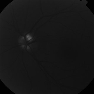
 Optic Disc Drusen
Apr 2 2016 by David Callanan, MD
30-year-old Caucasian male with visual field defect OD > OS.
Condition/keywords: optic disc drusen
-
 Autofluorescence of Optic Disc Drusen
Mar 2 2014 by Homayoun Tabandeh, MD, FASRS
Autofluorescence of optic disc drusen.
Condition/keywords: optic disc drusen
 Loading…