Search results (111 results)
-
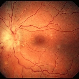 Toxoplasma Neuroretinitis (Jensen`s Disease)
Feb 25 2013 by Henry J. Kaplan, MD
Toxoplasma neuroretinitis in the left eye of a patient with macular star formation, retinitis adjacent to the optic nerve head with disc swelling.
Condition/keywords: Jensen disease, ocular toxoplasmosis, toxoplasmosis
-
 Ocular Toxocariasis slide 2
Oct 22 2012 by Ronald C. Gentile, MD
The fold of the retina was dry and cord like.
Photographer: The New York Eye & Ear Infirmary Department of Medical Imaging
Condition/keywords: ocular toxoplasmosis
-
 Toxocara Granuloma
Feb 25 2013 by Henry J. Kaplan, MD
Toxocara granuloma in the midperiphery of the retina.
Condition/keywords: ocular toxoplasmosis, toxocara granuloma, toxocariasis
-
 Ocular Toxoplasmosis Scar, Fluorescein Angiogram
Aug 23 2012 by Gerardo Garcia-Aguirre, MD
Fluorescein angiogram showing a large hypofluorescent round lesion with well-defined borders, where the fluorescence of the choroidal vessels is observed.
Photographer: Noemí Hernández, Asociación para Evitar la Ceguera en México
Imaging device: Zeiss FF4
Condition/keywords: toxoplasmosis
-
 Toxocara Granuloma
Feb 25 2013 by Henry J. Kaplan, MD
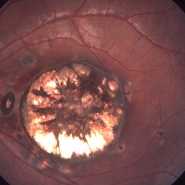
Toxocara granuloma superotemporal to the fovea.
Condition/keywords: ocular toxoplasmosis, toxocara granuloma, toxocariasis
-
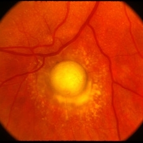
 Toxocara Granuloma
Feb 25 2013 by Henry J. Kaplan, MD
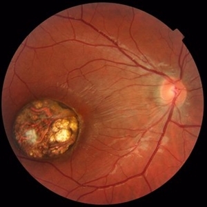
Toxocara granuloma of the optic nerve head.
Condition/keywords: ocular toxoplasmosis, toxocara granuloma, toxocariasis
-
 Toxoplasma chorioretinitis 2
Jan 11 2013 by Alex P. Hunyor, MD
Toxoplasmosis 2 - recurrent toxoplasma chorioretinitis at the margin of previous scar.
Condition/keywords: ocular toxoplasmosis, toxoplasmosis, toxoplasmosis retinitis
-
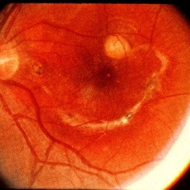
 Toxoplasma chorioretinitis 1
Jan 11 2013 by Alex P. Hunyor, MD
Toxoplasmosis 1 - chorioretinal scar from previous toxoplasma chorioretinitis. See image 2 - recurrent todo adjacent to this scar
Condition/keywords: inactive toxoplasmosis, ocular toxoplasmosis, toxoplasmosis, toxoplasmosis retinitis
-
---thumb.jpg/image-square;max$300,300.ImageHandler) Ocular Toxoplasmosis
Ocular Toxoplasmosis
Feb 15 2013 by From the Collections of Thomas M. Aaberg, MD and Thomas M. Aaberg Jr., MD
Diffuse slit-lamp photograph of the right eye of a patient with ocular toxoplasmosis showing strands of vitreous inflammation.
Condition/keywords: ocular toxoplasmosis, vitritis
-
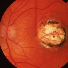
 Congenital Toxoplasmosis
Oct 10 2015 by Hamid Ahmadieh, MD
Color fundus photograph of the right eye of a 15 -year-old boy with decreased vision due to a large chorioretinal scar involving the macula . The lesion is typical for a congenital ocular toxoplasmosis .
Photographer: Solmaz Shahmohammad, Negah Eye Center, Tehran, Iran
Condition/keywords: color fundus photograph, congenital toxoplasmosis
-
 Toxocara Granuloma
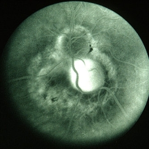
Feb 25 2013 by Henry J. Kaplan, MD
Toxocara granuloma of ON, late stage F/A. #3 Late hyperfluorescence in the granuloma due to staining.
Condition/keywords: ocular toxoplasmosis, toxocara granuloma, toxocariasis
-
 Ocular Toxoplasmosis
Nov 20 2015 by Ahmad B. Tarabishy, MD
28-year-old male with active toxoplasmosis chorioretinitis OS and a large macular toxoplasmosis scar OD. Vision is 20/25 OU.
Photographer: Phaedra Lund, Retina Specialists of Tampa
Imaging device: Zeiss Cirrus OCT
Condition/keywords: inactive toxoplasmosis, macula lesion, toxoplasmosis
-
 Ocular Toxoplasmosis Scar, Fundus Photograph
Aug 23 2012 by Gerardo Garcia-Aguirre, MD
Fundus photograph showing a large round atrophic lesion in the posterior pole, with pigment migration and several satellite lesions.
Photographer: Noemí Hernández, Asociación para Evitar la Ceguera en México
Imaging device: Zeiss FF4
-
 Ocular Toxoplasmosis
Nov 20 2015 by Ahmad B. Tarabishy, MD
28-year-old male with active toxoplasmosis chorioretinitis OS and a large macular toxoplasmosis scar OD. Vision is 20/25 OU.
Photographer: Phaedra Lund, Retina Specialists of Tampa
Imaging device: Zeiss Cirrus OCT
Condition/keywords: acute toxoplasmosis, toxoplasmosis, toxoplasmosis chorioretinitis, toxoplasmosis reactivation
-
---thumb.jpg/image-square;max$300,300.ImageHandler) vitreous haze and retinal detachment
vitreous haze and retinal detachment
Feb 14 2013 by From the Collections of Thomas M. Aaberg, MD and Thomas M. Aaberg Jr., MD
color fundus photograph showing vitreous haze and retinal detachment associated with ocular toxoplasmosis.
Condition/keywords: exudative retinal detachment, ocular toxoplasmosis
-
---thumb.jpg/image-square;max$300,300.ImageHandler) vitreous haze and retinal detachment associated with ocular toxoplasmosis
vitreous haze and retinal detachment associated with ocular toxoplasmosis
Feb 14 2013 by From the Collections of Thomas M. Aaberg, MD and Thomas M. Aaberg Jr., MD
color fundus photograph showing vitreous haze and retinal detachment associated with ocular toxoplasmosis
Condition/keywords: exudative retinal detachment, ocular toxoplasmosis
-
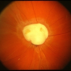
 Toxocara Granuloma
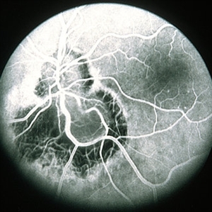
Feb 25 2013 by Henry J. Kaplan, MD
Toxocara granuloma of optic nerve; F/A.
Condition/keywords: ocular toxoplasmosis, toxocara granuloma, toxocariasis
-
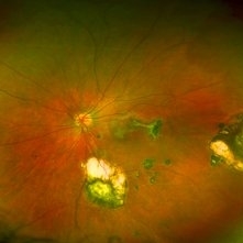 Toxoplasmosis
Jun 3 2017 by Gabriel Costa Andrade, PhD
Fundus photograph of an 14-year-old boy with multiple chorioretinal scars secondary to toxoplasmosis.
Photographer: Gabriel Costa de Andrade
Imaging device: Optos® California
Condition/keywords: congenital toxoplasmosis, ocular toxoplasmosis, toxoplasmosis chorioretinitis, toxoplasmosis uveitis
-
---thumb.jpg/image-square;max$300,300.ImageHandler) vitreous haze and confluent peripheral retinal whitening consistent with active ocular toxoplasmosis
vitreous haze and confluent peripheral retinal whitening consistent with active ocular toxoplasmosis
Feb 15 2013 by From the Collections of Thomas M. Aaberg, MD and Thomas M. Aaberg Jr., MD
Color fundus photograph showing vitreous haze and confluent peripheral retinal whitening consistent with active ocular toxoplasmosis
Condition/keywords: ocular toxoplasmosis, vitritis
-
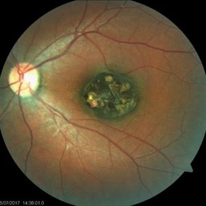 Congenital Toxoplasmosis
Jul 22 2017 by Akif Erol
Color fundus photograph of the left eye of an 18-year-old girl with decreased vision due to a large chorioretinal scar involving the macula. The lesion is typical for a congenital ocular toxoplasmosis
Photographer: Mehmet Akif Erol, Afyon Kocatepe University Ophthalmology Clinic
Condition/keywords: color fundus photograph, congenital toxoplasmosis
-
---thumb.jpg/image-square;max$300,300.ImageHandler) Vitreous haze and confluent peripheral retinal whitening
Vitreous haze and confluent peripheral retinal whitening
Feb 15 2013 by From the Collections of Thomas M. Aaberg, MD and Thomas M. Aaberg Jr., MD
Color fundus photograph showing vitreous haze and confluent peripheral retinal whitening consistent with active ocular toxoplasmosis.
Condition/keywords: ocular toxoplasmosis, vitritis
-
---thumb.jpg/image-square;max$300,300.ImageHandler) Foci of arteriolar plaques
Foci of arteriolar plaques
Feb 15 2013 by From the Collections of Thomas M. Aaberg, MD and Thomas M. Aaberg Jr., MD
color fundus photograph showing foci of arteriolar plaques (so-called Kyrieleis arteritis), as seen in ocular toxoplasmosis.
Condition/keywords: ocular toxoplasmosis
-
 Congenital Toxoplasmosis
Apr 8 2019 by Gary R. Cook, MD, FACS
Left eye of the same 38-year-old female with congenital toxoplasmosis lesion; V.A. = 20/40 due to temporal location of the Toxo scar.
Imaging device: Topcon VT-50
Condition/keywords: chorioretinal scar, congenital toxoplasmosis, inactive toxoplasmosis, macular scar, ocular toxoplasmosis
-
---thumb.jpg/image-square;max$300,300.ImageHandler) Vitreous haze and focal peripheral retinal whitening consistent with active ocular toxoplasmosis
Vitreous haze and focal peripheral retinal whitening consistent with active ocular toxoplasmosis
Feb 15 2013 by From the Collections of Thomas M. Aaberg, MD and Thomas M. Aaberg Jr., MD
Color fundus photograph showing vitreous haze and focal peripheral retinal whitening consistent with active ocular toxoplasmosis
Condition/keywords: ocular toxoplasmosis, vitritis
-
---thumb.jpg/image-square;max$300,300.ImageHandler) Vitreous haze, disc edema and indistinct foci of retinal whitening
Vitreous haze, disc edema and indistinct foci of retinal whitening
Feb 15 2013 by From the Collections of Thomas M. Aaberg, MD and Thomas M. Aaberg Jr., MD
Color fundus photograph showing vitreous haze, disc edema and indistinct foci of retinal whitening consistent with active ocular toxoplasmosis.
Condition/keywords: ocular toxoplasmosis, vitritis
 Loading…