Search results (65 results)
-
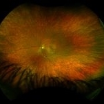
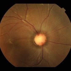
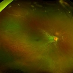
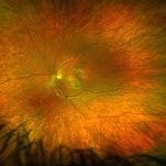 "Mud-Splatter" of Posterior Pole and Peripheral Radial Streaks in a Carrier of Ocular Albinism
Jan 22 2019 by John S. King, MD
14-year-old healthy white female with family history of ocular albinism was seen by Dr. Hruby for a second opinion. Father and some of his brothers were positive for a history of ocular albinism. Va cc 20/30 J1+ OU; no nystagmus; no TIDs; no foveal hypoplasia. A "mud-spatter" appearance to the posterior pole was present, along with peripheral alternating streaks (photo). Dr. Hruby agreed that this was most likely a carrier of Ocular Albinism Type-1 (XR; GPR143 mutation), and possible genetic testing/counselling was discussed.
Photographer: Gretchen Harper
Imaging device: Optos California
Condition/keywords: Nettleship-Falls ocular albinism, ocular albinism
-
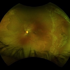
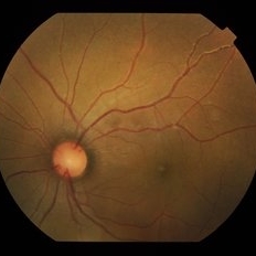
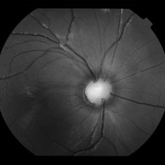
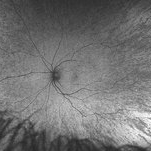 "Mud-Splatter" of Posterior Pole and Peripheral Radial Streaks in a Carrier of Ocular Albinism
Jan 22 2019 by John S. King, MD
14-year-old healthy white female with family history of ocular albinism was seen by Dr. Hruby for a second opinion. Father and some of his brothers were positive for a history of ocular albinism. Va cc 20/30 J1+ OU; no nystagmus; no TIDs; no foveal hypoplasia. A "mud-spatter" appearance to the posterior pole was present, along with peripheral alternating streaks. Hypoautofluorescent areas correspond to hyperpigmented areas of retinal pigment epithelium, and vice versa (see photo). Dr. Hruby agreed that this was most likely a carrier of Ocular Albinism Type-1 (XR; GPR143 mutation), and possible genetic testing/counselling was discussed.
Photographer: Gretchen Harper
Imaging device: Optos California
Condition/keywords: Nettleship-Falls ocular albinism, ocular albinism
-
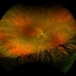 "Mud-Splatter" of Posterior Pole and Peripheral Radial Streaks in a Carrier of Ocular Albinism
Jan 22 2019 by John S. King, MD
14-year-old healthy white female with family history of ocular albinism was seen by Dr. Hruby for a second opinion. Father and some of his brothers were positive for a history of ocular albinism. Va cc 20/30 J1+ OU; no nystagmus; no TIDs; no foveal hypoplasia. A "mud-spatter" appearance to the posterior pole was present, along with peripheral alternating streaks (photo). Dr. Hruby agreed that this was most likely a carrier of Ocular Albinism Type-1 (XR; GPR143 mutation), and possible genetic testing/counselling was discussed.
Photographer: Gretchen Harper
Imaging device: Optos California
Condition/keywords: Nettleship-Falls ocular albinism, ocular albinism
-
 Progressive Bifocal Chorioretinal Atrophy
Feb 1 2015 by Andree Henaine-Berra, MD
Fundus photograph of the left eye of an 13-year-old female patient with poor vision, high myopia and nystagmus. The image shows macular dragging, a limited area of chorioretinal atrophy temporal to the optic disc and an extense area of chorioretinal atrophy temporal to the macula that extended to the extreme periphery.
Photographer: Andree Henaine-Berra, MD
Condition/keywords: chorioretinal atrophy
-
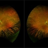 "Mud-Splatter" of Posterior Pole and Peripheral Radial Streaks in a Carrier of Ocular Albinism
Jan 22 2019 by John S. King, MD
14-year-old healthy white female with family history of ocular albinism was seen by Dr. Hruby for a second opinion. Father and some of his brothers were positive for a history of ocular albinism. Va cc 20/30 J1+ OU; no nystagmus; no TIDs; no foveal hypoplasia. A "mud-spatter" appearance to the posterior pole was present, along with peripheral alternating streaks (photo). Dr. Hruby agreed that this was most likely a carrier of Ocular Albinism Type-1 (XR; GPR143 mutation), and possible genetic testing/counselling was discussed.
Photographer: Gretchen Harper
Imaging device: Optos California
Condition/keywords: Nettleship-Falls ocular albinism, ocular albinism
-
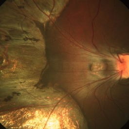
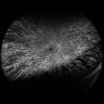 "Mud-Splatter" of Posterior Pole and Peripheral Radial Streaks in a Carrier of Ocular Albinism
Jan 22 2019 by John S. King, MD
14-year-old healthy white female with family history of ocular albinism was seen by Dr. Hruby for a second opinion. Father and some of his brothers were positive for a history of ocular albinism. Va cc 20/30 J1+ OU; no nystagmus; no TIDs; no foveal hypoplasia. A "mud-spatter" appearance to the posterior pole was present, along with peripheral alternating streaks, which are very prominent in this late phase FA of the right eye. Dr. Hruby agreed that this was most likely a carrier of Ocular Albinism Type-1 (XR; GPR143 mutation), and possible genetic testing/counselling was discussed.
Photographer: Gretchen Harper
Imaging device: Optos California
Condition/keywords: Nettleship-Falls ocular albinism, ocular albinism
-
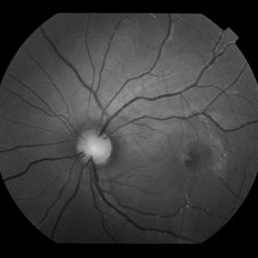
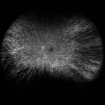 "Mud-Splatter" of Posterior Pole and Peripheral Radial Streaks in a Carrier of Ocular Albinism
Jan 22 2019 by John S. King, MD
14-year-old healthy white female with family history of ocular albinism was seen by Dr. Hruby for a second opinion. Father and some of his brothers were positive for a history of ocular albinism. Va cc 20/30 J1+ OU; no nystagmus; no TIDs; no foveal hypoplasia. A "mud-spatter" appearance to the posterior pole was present, along with peripheral alternating streaks that are very prominent on this late phase FA OS. Dr. Hruby agreed that this was most likely a carrier of Ocular Albinism Type-1 (XR; GPR143 mutation), and possible genetic testing/counselling was discussed.
Photographer: Gretchen Harper
Imaging device: Optos California
Condition/keywords: Nettleship-Falls ocular albinism, ocular albinism
-
 "Mud-Splatter" of Posterior Pole and Peripheral Radial Streaks in a Carrier of Ocular Albinism
Jan 22 2019 by John S. King, MD
14-year-old healthy white female with family history of ocular albinism was seen by Dr. Hruby for a second opinion. Father and some of his brothers were positive for a history of ocular albinism. Va cc 20/30 J1+ OU; no nystagmus; no TIDs; no foveal hypoplasia. A "mud-spatter" appearance to the posterior pole was present, along with peripheral alternating streaks (photo). Dr. Hruby agreed that this was most likely a carrier of Ocular Albinism Type-1 (XR; GPR143 mutation), and possible genetic testing/counselling was discussed.
Photographer: Gretchen Harper
Imaging device: Optos California
Condition/keywords: Nettleship-Falls ocular albinism, ocular albinism
-
 Myopia with Lattice Degeneration and White Without Pressure in the Setting of Marfan's Syndrome
Aug 31 2020 by Sophia El Hamichi, MD
A 1-year-old female with Marfan's syndrome, myopia OU, congenital nystagmus and exotopia OD. Ultra-wide field imaging of her left eye showed lattice degeneration with atrophic retinal holes temporally, in addition to multiple sections of white without pressure.
Imaging device: Optos
Condition/keywords: atrophic retinal hole, lattice degeneration, Marfan's syndrome, myopia, Optos, ultra-wide field imaging
-
 Oculocutaneous albinism Slide 1
Oct 22 2012 by Ronald C. Gentile, MD
16-year-old girl with poor vision and nystagmus. She had oculocutaneous albinism with reduced pigmentation of the the iris with transillumination.
Photographer: The New York Eye & Ear Infirmary Department of Medical Imaging
Condition/keywords: ocular albinism
-
 Progressive Bifocal Chorioretinal Atrophy
Feb 1 2015 by Andree Henaine-Berra, MD
Fundus photograph of the right eye of an 13-year-old female patient with poor vision, high myopia and nystagmus. The image shows macular dragging, a limited area of chorioretinal atrophy temporal to the optic disc and an extense area of chorioretinal atrophy temporal to the macula that extended to the extreme periphery.
Photographer: Andree Henaine-Berra, MD
Condition/keywords: chorioretinal atrophy
-
 Progressive Bifocal Chorioretinal Atrophy
Feb 1 2015 by Andree Henaine-Berra, MD
Fluorescein angiography of the left eye of an 13-year-old female patient with poor vision, high myopia and nystagmus. The image shows a hypofluorescent area corresponding to the area of chorioretinal atrophy. Some middle sized choroidal vessels can still be observed.
Photographer: Andree Henaine-Berra, MD
Condition/keywords: chorioretinal atrophy
-
 Optic Disc Coloboma
Jul 24 2019 by Haider Ali
16-year-old boy with horizontal nystagmus and decreased vision in both eyes.
Photographer: Dr Haider Ali Chaudhry, Madinah Teaching Hospital, Faisalabad
Condition/keywords: coloboma, coloboma of optic disc, coloboma of the optic nerve, excavation, Morning Glory Syndrome
-
 Progressive Bifocal Corioretinal Atrophy
Feb 1 2015 by Andree Henaine-Berra, MD
Fluorescein angiography of the right eye of an 13-year-old female patient with poor vision, high myopia and nystagmus. The image shows a hypofluorescent area corresponding to the area of chorioretinal atrophy. Some middle sized choroidal vessels can still be observed.
Photographer: Andree Henaine-Berra, MD
Condition/keywords: chorioretinal atrophy
-
 Optic Disc Coloboma
Jul 24 2019 by Haider Ali
16-year-old boy with horizontal nystagmus and decreased vision in both eyes.
Photographer: Dr Haider Ali Chaudhry, Madinah Teaching Hospital, Faisalabad
Condition/keywords: coloboma, coloboma of optic disc, coloboma of the optic nerve, excavation, Morning Glory Syndrome
-
 Retinopathy of Prematurity
Oct 8 2019 by Olivia Rainey
Ultra-wide field pseudocolor image of a 15-year-old male with retinopathy of prematurity affecting both of his eyes. Patient was born at 22 weeks and had a birth weight of 434g. He presented with esotropia, nystagmus, and severe macular/vascular dragging in the right eye.
Photographer: Olivia Rainey
Imaging device: Optos
Condition/keywords: esotropia, macular dragging, myopia, nystagmus, Optos, retinopathy of prematurity (ROP), ultra-wide field imaging, vascular arrest, vascular dragging
-
 Macular Coloboma With Macular Dystrophy
Jan 24 2020 by Deepak Bhojwani, MS
Fundus image of a 22-year-old gentlemen with poor vision since childhood with nystagmus. fundus photo showing macular coloboma which is rarely seen in macular dystrophies.
Photographer: DEEPAK BHOJWANI
Condition/keywords: macular dystrophy
-
 Macular Coloboma With Macular Dystrophy
Jan 24 2020 by Deepak Bhojwani, MS
Fundus image of a 22-year-old gentlemen with poor vision since childhood with nystagmus. Fundus photo showing macular coloboma which is rarely seen in macular dystrophies.
Photographer: DEEPAK BHOJWANI
Condition/keywords: coloboma of macula, macular dystrophy
-
 Optic Disc Coloboma
Jul 24 2019 by Haider Ali
16-year-old boy with horizontal nystagmus and decreased vision in both eyes.
Photographer: Dr Haider Ali Chaudhry, Madinah Teaching Hospital, Faisalabad
Condition/keywords: coloboma, coloboma of optic disc, coloboma of the optic nerve, excavation, Morning Glory Syndrome
-
 Achromatopsia, right eye
Oct 28 2019 by Albert Li, MD, FASRS
Fundus photograph of 38-year-old man with congenital horizontal nystagmus.
Condition/keywords: achromatopsia
-
 Oculocutaneous Albinism
Oct 24 2020 by Guilherme Daher
Oculocutaneous albinism is a group of rare inherited disorders characterized by a reduction or complete lack of melanin pigment in the skin, hair and eyes.
Photographer: Jefferson Rocha, Instituto Suel Abujamra, Sao Paulo Brazil
Condition/keywords: albinism, nystagmus, oculocutaneous albinism
-
 Flat Fovea in Oculocutaneous Albinism
Oct 24 2020 by Guilherme Daher
Optical coherence tomography of a patient with oculocutaneous albinism showing a flat fovea.
Photographer: Jefferson Rocha, Instituto Suel Abujamra, Sao Paulo Brazil
Imaging device: Zeiss Cirrus HD-OCT 5000
Condition/keywords: albinism, fovea, foveal hypoplasia, nystagmus, oculocutaneous albinism, optical coherence tomography (OCT)
-
 OCT of Achromatopsia, Right Eye
Oct 28 2019 by Albert Li, MD, FASRS
OCT of 38-year-old man with congenital horizontal nystagmus with central ellipsoid zone loss.
Imaging device: Heidelberg Spectralis
Condition/keywords: achromatopsia
-
 Iris in Albinism
May 1 2020 by Anfisa Ayalon, MD
Slit-lamp photograph of an 34-year-old man with oculocutaneous albinism. Note iris transillumination defects. Snellen chart visual acuity in both eyes-6/60. Nystagmus is also present.
Photographer: Anfisa Ayalon, MD., Meir Medical Center, Kfar Saba, Israel.
Condition/keywords: iris, nystagmus, oculocutaneous albinism, transillumination
 Loading…