Search results (65 results)
-
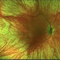
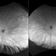
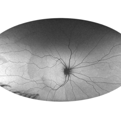 Foveal Hypoplasia AF
Feb 1 2025 by Poornachandra B, MS, FVRS
This is a wide field autofluorescence image of 21 year-old male. He presented with history of low vision since childhood associated with nystagmus. Uniform fluorescence across posterior pole with absent foveal hypo autofluorescence can be seen on the image.
Photographer: Mr Dhikshith
Condition/keywords: autofluorescence imaging, foveal hypoplasia, nystagmus, ultra-wide field imaging
-
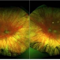
 Retinitis Pigmentosa with PPRPE - FAF-G
Jan 27 2025 by Vishal Agrawal, MD, FRCS,FACS,FASRS
16 year-old male patient presented with DOV, nyctalopia and nystagmus. Fundus revealed pigment clumping, pale disc and preserved para-arteriolar retinal pigment epithelium (PPRPE) in both eyes. Genetic testing revealed CRB1 gene mutation.
Photographer: Dr Ayushi Gupta
Imaging device: Clarus 700
Condition/keywords: retinitis pigmentosa
-
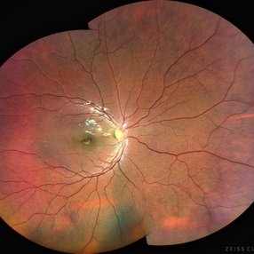
 Retinitis Pigmentosa with PPRPE
Jan 27 2025 by Vishal Agrawal, MD, FRCS,FACS,FASRS
16 year-old male patient presented with DOV, nyctalopia and nystagmus. Fundus revealed pigment clumping, pale disc and preserved para-arteriolar retinal pigment epithelium (PPRPE) in both eyes. Genetic testing revealed CRB1 gene mutation.
Photographer: Dr Ayushi
Imaging device: Clarus 700
Condition/keywords: retinitis pigmentosa
-
 Unilateral Coloboma Involving Disc and Macula
Dec 27 2024 by Tejaswita Verma
Fundus image of a 15 years old male presenting with unilaterally diminished vision since childhood in RE with CF3mt vision and inferior iris coloboma and retinochoroidal coloboma with nystagmus and cataract.
Photographer: DR. TEJASWITA VERMA
Imaging device: MIRANTE
Condition/keywords: chorioretinal coloboma, iridofundal coloboma
-
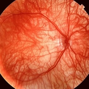 Foveal Hypoplasia / Ocular Albinism
Aug 29 2024 by César Adrián Gómez Valdivia, MD
Fundus photograph of a 6-year-old female patient with foveal hypoplasia, ocular albinism and pendular nystagmus. Findings were bilateral. Retinal and choroidal vasculature are exquisitely beautiful.
Photographer: @eyemissu2
Imaging device: TOPCON TRC-50DX
Condition/keywords: foveal hypoplasia, ocular albinism
-
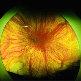 Foveal Hypoplasia / Ocular Albinism
Aug 29 2024 by César Adrián Gómez Valdivia, MD
Fundus photograph of a 64-year-old female patient with foveal hypoplasia, ocular albinism and pendular nystagmus. Findings were bilateral. Retinal and choroidal vasculature are exquisitely beautiful.
Photographer: @eyemissu2
Imaging device: California ICG OPTOS
Condition/keywords: foveal hypoplasia, ocular albinism
-
 Ocular Albinism
Aug 27 2024 by Monica Elena Cortizo Brown , MD
9 year old girl with pendular nystagmus, photophobia and a fundus photo that shows clear view of the choroidal vasculature due to the hypopigmentation of retinal pigment epithelium.
Photographer: Mónica Cortizo Brown , Hospital de la Luz, Ciudad de México
Condition/keywords: nystagmus, ocular albinism, oculocutaneous albinism, photophobia
-
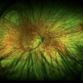 Foveal Hypoplasia / Ocular Albinism
Aug 25 2024 by César Adrián Gómez Valdivia, MD
Fundus photograph of a 6-year-old female patient with foveal hypoplasia, ocular albinism and pendular nystagmus. Findings were bilateral. Retinal and Choroidal vasculature are exquisitely beautiful.
Photographer: @eyemissu2
Imaging device: TOPCON TRC-50DX
Condition/keywords: albinism, foveal hypoplasia, ocular albinism
-
 Foveal Hypoplasia / Ocular Albinism
Aug 25 2024 by César Adrián Gómez Valdivia, MD
Fundus photograph of a 6-year-old female patient with foveal hypoplasia, ocular albinism and pendular nystagmus. Findings were bilateral. Retinal and choroidal vasculature are exquisitely beautiful.
Photographer: @eyemissu2
Imaging device: TOPCON TRC-50DX
Condition/keywords: Albinism, foveal hypoplasia, ocular albinism, vascula
-
 Oculocutaneous Albinism
Jan 14 2024 by Hemanth Murthy, MBBS, MD, FASRS
9 year boy presented with pendular nystagmus and blurring of vision. He light skin colour with light coloured hair. Fundus picture of right eye
Photographer: Mr Veda Vyas
Imaging device: Optos Daytona
Condition/keywords: albinism
-
 Oculocutaneous Albinism
Jan 14 2024 by Hemanth Murthy, MBBS, MD, FASRS
Fundus image of left eye showing near absence of pigment in a 9 year boy with blurring of vision and pendular nystagmus. The skin colour and hair was light coloured and OCT showed foveal hypoplasia
Photographer: Mr Veda Vyas
Imaging device: Optos Daytona
Condition/keywords: Albinism
-
 Cone Dystrophy
Aug 30 2023 by Vishal Agrawal, MD, FRCS,FACS,FASRS
12 year old male patient presented with photophobia, decrease in vision and Nystagmus. Bulls eye maculopathy gives an appearance of an eye on the fovea on color fundus photo due to nystagmus.
Photographer: Dr Bhagyashree
Imaging device: Clarus 700
Condition/keywords: bull's eye maculopathy, Cone-Rod Dystrophy
-
 Ocular albinism
Aug 18 2023 by Dr.Anushri Godbole
41 years old male came to opd with chief complaints of diminution of vision of both eyes since childhood. BCVA RE- FC 2M, LE- 6/60. On examination, patient had BE nystagmus. Fundus was tessellated with prominent choroidal blood vessels and Foveolar hypoplasia. Diagnosis of BE ocular albinism was made..
Condition/keywords: ocular albinism
-
 Ocular albinism
Aug 18 2023 by Dr.Anushri Godbole
41 years old male came to opd with chief complaints of diminution of vision of both eyes since childhood. BCVA RE- FC 2M, LE- 6/60. On examination, Patient had BE nystagmus. Fundus was tessellated with prominent choroidal blood vessels and Foveolar hypoplasia. Diagnosis of BE ocular albinism was made..
Condition/keywords: ocular albinism
-
 Ocular Albinism
Apr 10 2023 by William Jacob Anderson, MD
Fundus photograph of a 21-year-old girl with ocular albinism. Clinical exam was significant for decreased visual acuity and nystagmus. OCT macula demonstrates foveal hypoplasia
Photographer: William J Anderson, MD, Saint Louis University
Imaging device: Heidelberg Spectralis OCT
Condition/keywords: Albinism, foveal hypoplasia, ocular albinism
-
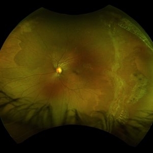
 Oculocutaneous Albinism
Jan 22 2023 by Pietro Dechichi
Fundus photograph of an 6-year-old girl with oculocutaneous albinism. Pacient´s nystagmus made it difficult to perform the exame. Foveal hypoplasia and evident choroidal vessels can be seen in the retinography
Photographer: Pietro Dechichi
Imaging device: Optos California
Condition/keywords: childhood, foveal hypoplasia, ocular albinism
-
 Oculocutaneous Albinism
Jan 22 2023 by Pietro Dechichi
Fundus photograph of a 6-year-old girl with oculocutaneous albinism. Patient's nystagmus made it difficult to perform the exam. Foveal hypoplasia and evident choroidal vessels can be seen in the retinography
Photographer: Pietro Dechichi
Imaging device: Optos California
Condition/keywords: childhood, foveal hypoplasia, ocular albinism
-
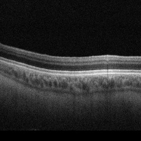
 Flat Fovea in Oculocutaneous Albinism
Oct 24 2020 by Guilherme Daher
Optical coherence tomography of a patient with oculocutaneous albinism showing a flat fovea.
Photographer: Jefferson Rocha, Instituto Suel Abujamra, Sao Paulo Brazil
Imaging device: Zeiss Cirrus HD-OCT 5000
Condition/keywords: albinism, fovea, foveal hypoplasia, nystagmus, oculocutaneous albinism, optical coherence tomography (OCT)
-
 Oculocutaneous Albinism Fundus Photograph
Oct 24 2020 by Guilherme Daher
Foveal hypoplasia in a oculocutaneous albinism patient. Who also has nystagmus associated.
Photographer: Jefferson Rocha, Instituto Suel Abujamra, Sao Paulo Brazil
Condition/keywords: albinism, fovea, foveal hypoplasia, hypoplasia, nystagmus, oculocutaneous albinism
-
 Oculocutaneous Albinism
Oct 24 2020 by Guilherme Daher
Oculocutaneous albinism is a group of rare inherited disorders characterized by a reduction or complete lack of melanin pigment in the skin, hair and eyes.
Photographer: Jefferson Rocha, Instituto Suel Abujamra, Sao Paulo Brazil
Condition/keywords: albinism, nystagmus, oculocutaneous albinism
-
 Myopia with Lattice Degeneration and White Without Pressure in the Setting of Marfan's Syndrome
Aug 31 2020 by Sophia El Hamichi, MD
A 1-year-old female with Marfan's syndrome, myopia OU, congenital nystagmus and exotopia OD. Ultra-wide field imaging of her left eye showed lattice degeneration with atrophic retinal holes temporally, in addition to multiple sections of white without pressure.
Imaging device: Optos
Condition/keywords: atrophic retinal hole, lattice degeneration, Marfan's syndrome, myopia, Optos, ultra-wide field imaging
-
 Lamellar Congenital Cataracts
Aug 24 2020 by Sophia El Hamichi, MD
A 4-month-old female with family history of congenital cataract was referred for bilateral congenital cataract with nystagmus and torticoli. During the ophthalmological exam we were able to access the fundus and perform a fluorescein angiogram through the clear ring surrounding the lens opacity. Retina evaluation showed no abnormalities.
Photographer: Abby Orcutt-Hayes, Murray Ocular Oncology and Retina
Imaging device: RetCam
Condition/keywords: congenital cataract, lamellar cataract, nystagmus
-
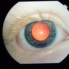
 Iris in Albinism
May 1 2020 by Anfisa Ayalon, MD
Slit-lamp photograph of an 34-year-old man with oculocutaneous albinism. Note iris transillumination defects. Snellen chart visual acuity in both eyes-6/60. Nystagmus is also present.
Photographer: Anfisa Ayalon, MD., Meir Medical Center, Kfar Saba, Israel.
Condition/keywords: iris, nystagmus, oculocutaneous albinism, transillumination
-
 Macular Coloboma With Macular Dystrophy
Jan 24 2020 by Deepak Bhojwani, MS
Fundus image of a 22-year-old gentlemen with poor vision since childhood with nystagmus. Fundus photo showing macular coloboma which is rarely seen in macular dystrophies.
Photographer: DEEPAK BHOJWANI
Condition/keywords: coloboma of macula, macular dystrophy
 Loading…